Effect of Patient Characteristics on Vessel Enhancement at Lower Extremity CT Angiography
- Affiliations
-
- 1Department of Radiological Technology, Tsuchiya General Hospital, Hiroshima 730-8655, Japan. takanorimasuda@yahoo.co.jp
- 2Department of Diagnostic Radiology, Tsuchiya General Hospital, Hiroshima 730-8655, Japan.
- 3Department of Diagnostic Radiology, Graduate School of Medical Sciences, Kumamoto University, Kumamoto 860-8556, Japan.
- 4Department of Medical Physics, Faculty of Life Sciences, Kumamoto University, Kumamoto 860-8556, Japan.
- 5Department of Diagnostic Radiology, Graduate School of Biomedical Sciences, Hiroshima University, Hiroshima 734-0037, Japan.
- KMID: 2404923
- DOI: http://doi.org/10.3348/kjr.2018.19.2.265
Abstract
OBJECTIVE
To evaluate the effect of patient characteristics on popliteal aortic contrast enhancement at lower extremity CT angiography (LE-CTA) scanning.
MATERIALS AND METHODS
Prior informed consent to participate was obtained from all 158 patients. All were examined using a routine protocol; the scanning parameters were tube voltage 100 kVp, tube current 100 mA to 770 mA (noise index 12), 0.5-second rotation, 1.25-mm detector row width, 0.516 beam pitch, and 41.2-mm table movement, and the contrast material was 85.0 mL. Cardiac output (CO) was measured with a portable electrical velocimeter within 5 minutes of starting the CT scan. To evaluate the effects of age, sex, body size, CO, and scan delay on the CT number of popliteal artery, the researchers used multivariate regression analysis.
RESULTS
A significant positive correlation was seen between the CT number of the popliteal artery and the patient age (r = 0.39, p < 0.01). A significant inverse correlation was observed between the CT number of the popliteal artery and the height (r = −0.48), total body weight (r = −0.52), body mass index (r = −0.33), body surface area (BSA) (r = −0.56), lean body weight (r = −0.56), and CO (r = −0.35) (p < 0.001 for all). There was no significant correlation between the enhancement and the scan delay (r = 0.06, p = 0.47). The BSA, CO, and age had significant effects on the CT number (standardized regression: BSA −0.42, CO −0.22, age 0.15; p < 0.05, respectively).
CONCLUSION
The BSA, CO, and age are significantly correlated with the CT number of the popliteal artery on LE-CTA.
MeSH Terms
Figure
-

Fig. 1 CT attenuation measurement in popliteal artery.A. Volume-rendering image of at LE-CTA in 77-year-old man. B. Maximum-intensity projection at LE-CTA. C. Measurement sites of volume rendering image for popliteal arteries at level of patella. D. Measurement sites of axial image for popliteal arteries at level of patella. CT = computed tomography, LE-CTA = lower extremity CT angiography
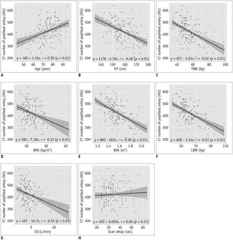
Fig. 2 Relationship between popliteal arterial enhancement and patient characteristics.Scattergrams of relationship between arterial enhancement obtained with protocol, using fixed dose of iodinated contrast material and patient age (A), HT (B), TBW (C), BMI (D), BSA (E), LBW (F), CO (G), and scan delay (H). There was significant positive correlation between CT number of popliteal artery and age (r = 0.39, p < 0.01). Correlation was inverse with HT (r = −0.48), TBW (r = −0.52), BMI (r = −0.33), BSA (r = −0.56), LBW (r = −0.53), and CO (r = −0.35) by linear regression analysis (p < 0.01 for all). There was no significant correlation between vessel enhancement and scan delay (r = 0.06, p = 0.47). BMI = body mass index, BSA = body surface area, CO = cardiac output, HT = height, HU = Hounsfield units, LBW = lean body weight, TBW = total body weight
Reference
-
1. Criqui MH, Fronek A, Barrett-Connor E, Klauber MR, Gabriel S, Goodman D. The prevalence of peripheral arterial disease in a defined population. Circulation. 1985; 71:510–515. PMID: 3156006.
Article2. Schroll M, Munck O. Estimation of peripheral arteriosclerotic disease by ankle blood pressure measurements in a population study of 60-year-old men and women. J Chronic Dis. 1981; 34:261–269. PMID: 7240365.
Article3. Selvin E, Erlinger TP. Prevalence of and risk factors for peripheral arterial disease in the United States: results from the National Health and Nutrition Examination Survey, 1999-2000. Circulation. 2004; 110:738–743. PMID: 15262830.
Article4. Chetter IC, Dolan P, Spark JI, Scott DJ, Kester RC. Correlating clinical indicators of lower-limb ischaemia with quality of life. Cardiovasc Surg. 1997; 5:361–366. PMID: 9350789.
Article5. Bloor K. Natural history of arteriosclerosis of the lower extremities: Hunterian lecture delivered at the Royal College of surgeons of England on 22nd April 1960. Ann R Coll Surg Engl. 1961; 28:36–52. PMID: 19310276.6. Dormandy J, Heeck L, Vig S. The fate of patients with critical leg ischemia. Semin Vasc Surg. 1999; 12:142–147. PMID: 10777241.7. Adam DJ, Beard JD, Cleveland T, Bell J, Bradbury AW, Forbes JF, et al. Bypass versus angioplasty in severe ischaemia of the leg (BASIL): multicentre, randomised controlled trial. Lancet. 2005; 366:1925–1934. PMID: 16325694.
Article8. Norgren L, Hiatt WR, Dormandy JA, Nehler MR, Harris KA, Fowkes FG. TASC II Working Group. Inter-Society Consensus for the Management of Peripheral Arterial Disease (TASC II). J Vasc Surg. 2007; 45(Suppl S):S5–S67. PMID: 17223489.
Article9. Napoli A, Anzidei M, Zaccagna F, Cavallo Marincola B, Zini C, Brachetti G, et al. Peripheral arterial occlusive disease: diagnostic performance and effect on therapeutic management of 64-section CT angiography. Radiology. 2011; 261:976–986. PMID: 21969664.
Article10. Bae KT. Intravenous contrast medium administration and scan timing at CT: considerations and approaches. Radiology. 2010; 256:32–61. PMID: 20574084.
Article11. Schernthaner R, Stadler A, Lomoschitz F, Weber M, Fleischmann D, Lammer J, et al. Multidetector CT angiography in the assessment of peripheral arterial occlusive disease: accuracy in detecting the severity, number, and length of stenoses. Eur Radiol. 2008; 18:665–671. PMID: 18094974.
Article12. Pollak AW, Norton PT, Kramer CM. Multimodality imaging of lower extremity peripheral arterial disease: current role and future directions. Circ Cardiovasc Imaging. 2012; 5:797–807. PMID: 23169982.13. Yanaga Y, Awai K, Nakaura T, Utsunomiya D, Oda S, Hirai T, et al. Contrast material injection protocol with the dose adjusted to the body surface area for MDCT aortography. AJR Am J Roentgenol. 2010; 194:903–908. PMID: 20308489.
Article14. Boer P. Estimated lean body mass as an index for normalization of body fluid volumes in humans. Am J Physiol. 1984; 247(4 Pt 2):F632–F636. PMID: 6496691.
Article15. Mosteller RD. Simplified calculation of body-surface area. N Engl J Med. 1987; 317:1098. PMID: 3657876.
Article16. Hume R. Prediction of lean body mass from height and weight. J Clin Pathol. 1966; 19:389–391. PMID: 5929341.
Article17. Hallynck TH, Soep HH, Thomis JA, Boelaert J, Daneels R, Dettli L. Should clearance be normalised to body surface or to lean body mass? Br J Clin Pharmacol. 1981; 11:523–526. PMID: 7272167.
Article18. Itoh S, Ikeda M, Satake H, Ota T, Ishigaki T. The effect of patient age on contrast enhancement during CT of the pancreatobiliary region. AJR Am J Roentgenol. 2006; 187:505–510. PMID: 16861556.
Article19. Awai K, Kanematsu M, Kim T, Ichikawa T, Nakamura Y, Nakamoto A, et al. The optimal body size index with which to determine iodine dose for hepatic dynamic CT: a prospective multicenter study. Radiology. 2016; 278:773–781. PMID: 26356063.
Article20. Kohn JC, Lampi MC, Reinhart-King CA. Age-related vascular stiffening: causes and consequences. Front Genet. 2015; 6:112. PMID: 25926844.
Article21. Bae KT, Heiken JP, Brink JA. Aortic and hepatic contrast medium enhancement at CT. Part II. Effect of reduced cardiac output in a porcine model. Radiology. 1998; 207:657–662. PMID: 9609887.
Article
- Full Text Links
-
- Actions
-
Cited
- CITED
-
- Close
- Share
-
- Similar articles
-
- Is CT Angiography a Reliable Tool for Diagnosis of Traumatic Vessel Injury in the Lower Extremities?
- Preoperative Identification of Perforator Using CT Angiography in Fibular Osteocutaneous Free Flap Head and Neck Reconstruction
- Computed tomographic findings in posterior cranial fossa tumors: correlation between angiographic vascularityand CT enhancement
- Precedence of Parenchymal Enhancement on CT Angiography to a Fatal Duret Hemorrhage
- Adventitial Cystic Disease of Popliteal Artery
