Self-Gated Late Gadolinium Enhancement at 7T to Image Rats with Reperfused Acute Myocardial Infarction
- Affiliations
-
- 1Molecular Imaging Center, West China Hospital of Sichuan University, Chengdu 610041, China.
- 2Department of Radiology, West China Hospital of Sichuan University, Chengdu 610041, China. gaofabao@yahoo.com
- 3Mallinckrodt Institute of Radiology, Washington University School of Medicine in St. Louis, MO 63110, USA.
- KMID: 2404921
- DOI: http://doi.org/10.3348/kjr.2018.19.2.247
Abstract
OBJECTIVE
A failed electrocardiography (ECG)-trigger often leads to a long acquisition time (TA) and deterioration in image quality. The purpose of this study was to evaluate and optimize the technique of self-gated (SG) cardiovascular magnetic resonance (CMR) for cardiac late gadolinium enhancement (LGE) imaging of rats with myocardial infarction/reperfusion.
MATERIALS AND METHODS
Cardiovascular magnetic resonance images of 10 rats were obtained using SG-LGE or ECG with respiration double-gating (ECG-RESP-gating) method at 7T to compare differences in image interference and TA between the two methods. A variety of flip angles (FA: 10°-80°) and the number of repetitions (NR: 40, 80, 150, and 300) were investigated to determine optimal scan parameters of SG-LGE technique based on image quality score and contrast-to-noise ratio (CNR).
RESULTS
Self-gated late gadolinium enhancement allowed successful scan in 10 (100%) rats. However, only 4 (40%) rats were successfully scanned with the ECG-RESP-gating method. TAs with SG-LGE varied depending on NR used (TA: 41, 82, 154, and 307 seconds, corresponding to NR of 40, 80, 150, and 300, respectively). For the ECG-RESP-gating method, the average TA was 220 seconds. For SG-LGE images, CNR (42.5 ± 5.5, 43.5 ± 7.5, 54 ± 9, 59.5 ± 8.5, 56 ± 13, 54 ± 8, and 41 ± 9) and image quality score (1.85 ± 0.75, 2.20 ± 0.83, 2.85 ± 0.37, 3.85 ± 0.52, 2.8 ± 0.51, 2.45 ± 0.76, and 1.95 ± 0.60) were achieved with different FAs (10°, 15°, 20°, 25°, 30°, 35°, and 40°, respectively). Optimal FAs of 20°-30° and NR of 80 were recommended.
CONCLUSION
Self-gated technique can improve image quality of LGE without irregular ECG or respiration gating. Therefore, SG-LGE can be used an alternative method of ECG-RESP-gating.
Keyword
MeSH Terms
Figure
-
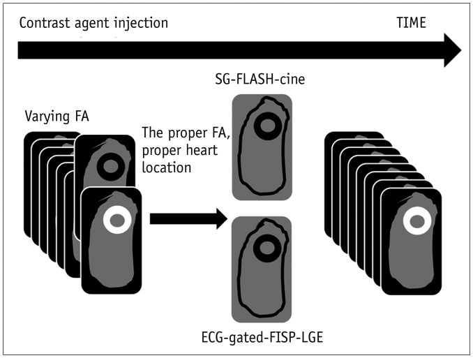
Fig. 1 Schematic summary of MRI protocol.Series of SG images were obtained after gadolinium injection, each with various FA. Last series of images with FAs of 20°–30° were used to select optimal FA images to yield best scar contrast. This FA was then used to perform subsequent SG FLASH imaging between 5–20 minutes after contrast agent administration. FISP-cine-Gate was then obtained following SG-FLASH-cine. ECG = electrocardiography, FA = flip angle, FISP = fast imaging with steady-state precession, FLASH = fast low angle shot MRI, LGE = late gadolinium enhancement, SG = self-gated
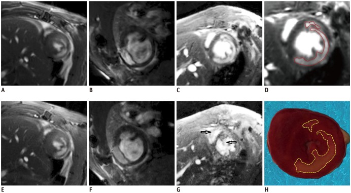
Fig. 2 SG-cine-LGE images (top) and ECG-gated-LGE images (bottom) are shown from three different rats.(A) and (E) are superb images after contrast agent injection whose scores were 4. (B) and (F) show a few artifacts that would not affect analysis results whose scores were 4. (C) and (G) were samples that ECG-gated technique could not show good enough images whose score was 1 while good images from SG sequence whose score was 3. (H) was triphenyl tetrazolium chloride staining demonstrating that infarction zone (in white or light pink color) was in anterior-lateral, lateral, and posterior-lateral wall of myocardium in (H), in good agreement with SG-LGE in (D). Black arrows (G) indicate flow and susceptibility artifacts in ECG-gated image which disappeared in SG image (C).
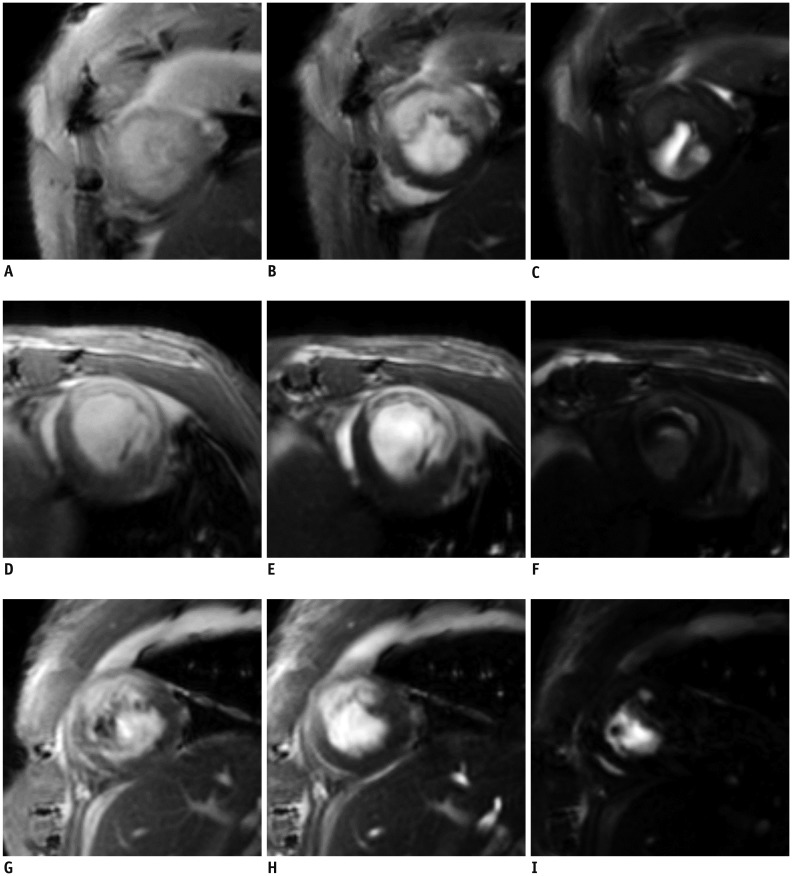
Fig. 3 Varying FA on LGE images are shown for three different rats.In left column, images (A, D, G) had FA of 10°. In middle column, images (B, E, H) had FAs of 20°–30°. In right column, images (C, F, I) had high FA of 60°–80°.
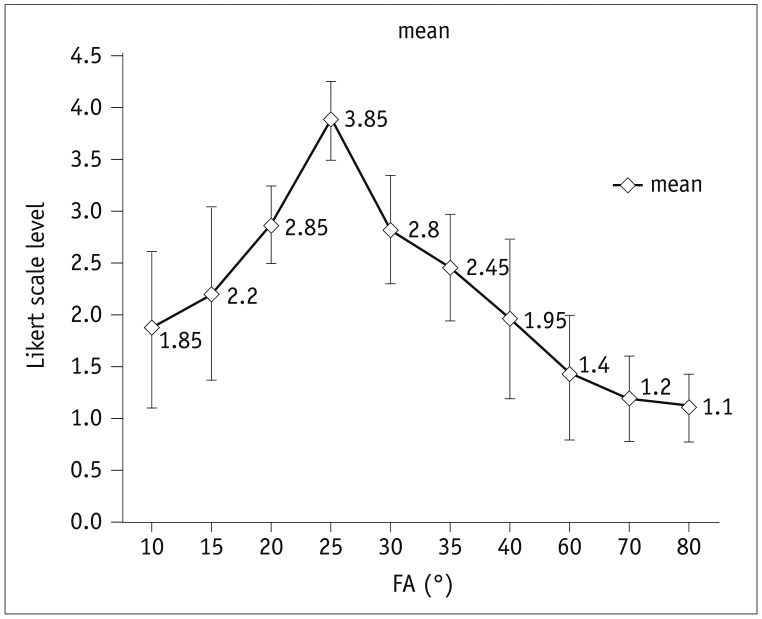
Fig. 4 Likert scale of SG sequence according to FAsQualitative grade was higher when FA was 20° to 30°. Qualities were minimized with lower or greater FA.
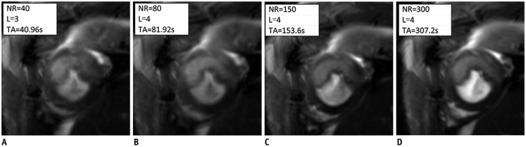
Fig. 5 Image quality and scan time varied with NR.(A-D) had NR of 40, 80, 150, and 300, respectively, with FA of 25°. Scan time was increased with increasing NR. NR = number of repetitions, L = Likert scale score, TA = acquisition time
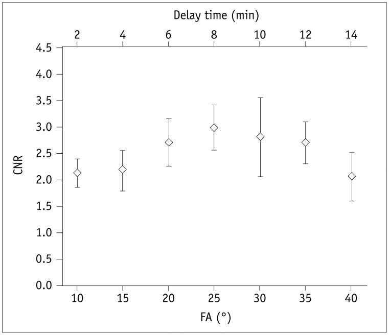
Fig. 6 CNR according to FA and delay time.Both delay time and FA were critical factors that affected CNRs of SG-LGE images. SG-LGE images were acquired within 2 minutes due to sequence preparation and TA for each SG scan. FA (bottom x-axis) was 10°, 15°, 20°, 25°, 30°, 35°, and 40°, corresponding to delay time (top x-axis) of 2, 4, 6, 8, 10, 12, and 14 minutes, respectively. CNR was increased rapidly to maximum values when FAs were 20° to 30° accompanied by delay time of 6 to 10 minutes. CNR = contrast-to-noise ratio
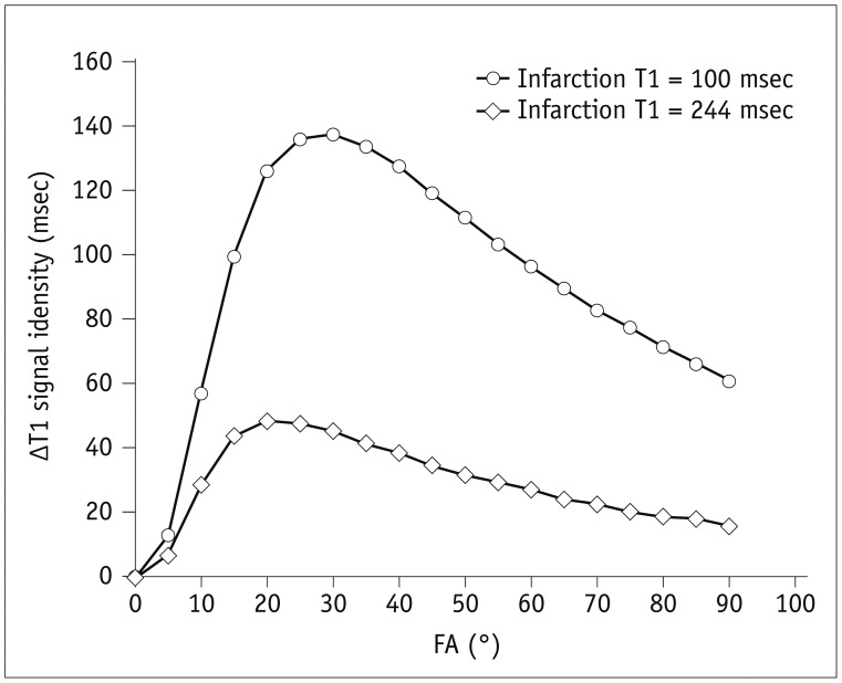
Fig. 7 Simulation of ΔT1 values under various FAs.Vertical axis ΔT1 values refer to deviations of T1 values between normal myocardium and infarction regions. They can be plotted as function of FA. Circle hollow plot: MI T1 = 100 msec; square hollow plot: MI T1 = 244 msec. MI = myocardial infarction
Cited by 2 articles
-
Guidelines for Cardiovascular Magnetic Resonance Imaging from the Korean Society of Cardiovascular Imaging—Part 2: Interpretation of Cine, Flow, and Angiography Data
Jae Wook Lee, Jee Hye Hur, Dong Hyun Yang, Bae Young Lee, Dong Jin Im, Su Jin Hong, Eun Young Kim, Eun-Ah Park, Yeseul Jo, JeongJae Kim, Chul Hwan Park, Hwan Seok Yong
Korean J Radiol. 2019;20(11):1477-1490. doi: 10.3348/kjr.2019.0407.Guideline for Cardiovascular Magnetic Resonance Imaging from the Korean Society of Cardiovascular Imaging—Part 1: Standardized Protocol
Yeseul Jo, JeongJae Kim, Chul Hwan Park, Jae Wook Lee, Jee Hye Hur, Dong Hyun Yang, Bae Young Lee, Dong Jin Im, Su Jin Hong, Eun Young Kim, Eun-Ah Park, Pan Ki Kim, Hwan Seok Yong
Korean J Radiol. 2019;20(9):1313-1333. doi: 10.3348/kjr.2019.0398.
Reference
-
1. Vandsburger MH, Epstein FH. Emerging MRI methods in translational cardiovascular research. J Cardiovasc Transl Res. 2011; 4:477–492. PMID: 21452060.
Article2. Abascal JF, Montesinos P, Marinetto E, Pascau J, Desco M. Comparison of total variation with a motion estimation based compressed sensing approach for self-gated cardiac cine MRI in small animal studies. PLoS One. 2014; 9:e110594. PMID: 25350290.
Article3. Paul J, Divkovic E, Wundrak S, Bernhardt P, Rottbauer W, Neumann H, et al. High-resolution respiratory self-gated golden angle cardiac MRI: comparison of self-gating methods in combination with k-t SPARSE SENSE. Magn Reson Med. 2015; 73:292–298. PMID: 24478142.
Article4. Yang Z, Berr SS, Gilson WD, Toufektsian MC, French BA. Simultaneous evaluation of infarct size and cardiac function in intact mice by contrast-enhanced cardiac magnetic resonance imaging reveals contractile dysfunction in noninfarcted regions early after myocardial infarction. Circulation. 2004; 109:1161–1167. PMID: 14967719.
Article5. Motaal AG, Noorman N, de Graaf WL, Hoerr V, Florack LM, Nicolay K, et al. Functional imaging of murine hearts using accelerated self-gated UTE cine MRI. Int J Cardiovasc Imaging. 2015; 31:83–94. PMID: 25204261.
Article6. Vallée JP, Ivancevic MK, Nguyen D, Morel DR, Jaconi M. Current status of cardiac MRI in small animals. MAGMA. 2004; 17:149–156. PMID: 15605278.
Article7. Berry CJ, Thedens DR, Light-McGroary K, Miller JD, Kutschke W, Zimmerman KA, et al. Effects of deep sedation or general anesthesia on cardiac function in mice undergoing cardiovascular magnetic resonance. J Cardiovasc Magn Reson. 2009; 11:16. PMID: 19454023.
Article8. Constantinides C, Mean R, Janssen BJ. Effects of isoflurane anesthesia on the cardiovascular function of the C57BL/6 mouse. ILAR J. 2011; 52:e21–e31. PMID: 21677360.9. Kramer K, van Acker SA, Voss HP, Grimbergen JA, van der, Bast A. Use of telemetry to record electrocardiogram and heart rate in freely moving mice. J Pharmacol Toxicol Methods. 1993; 30:209–215. PMID: 8123902.
Article10. Wang CC, Huang TY. Self-gated PROPELLER-encoded cine cardiac imaging. Int J Cardiovasc Imaging. 2012; 28:1477–1485. PMID: 22042429.
Article11. Berk WA, Shea MJ, Crevey BJ. Bradycardic responses to vagally mediated bedside maneuvers in healthy volunteers. Am J Med. 1991; 90:725–729. PMID: 2042688.
Article12. Kapa S, Venkatachalam KL, Asirvatham SJ. The autonomic nervous system in cardiac electrophysiology: an elegant interaction and emerging concepts. Cardiol Rev. 2010; 18:275–284. PMID: 20926936.13. Dimick RN, Hedlund LW, Herfkens RJ, Fram EK, Utz J. Optimizing electrocardiograph electrode placement for cardiac-gated magnetic resonance imaging. Invest Radiol. 1987; 22:17–22. PMID: 3818232.
Article14. Frauenrath T, Fuchs K, Dieringer MA, Özerdem C, Patel N, Renz W, et al. Detailing the use of magnetohydrodynamic effects for synchronization of MRI with the cardiac cycle: a feasibility study. J Magn Reson Imaging. 2012; 36:364–372. PMID: 22411274.
Article15. Polson MJ, Barker AT, Gardiner S. The effect of rapid rise-time magnetic fields on the ECG of the rat. Clin Phys Physiol Meas. 1982; 3:231–234. PMID: 7140162.
Article16. de Roquefeuil M, Vuissoz PA, Escanyé JM, Felblinger J. Effect of physiological heart rate variability on quantitative T2 measurement with ECG-gated Fast Spin Echo (FSE) sequence and its retrospective correction. Magn Reson Imaging. 2013; 31:1559–1566. PMID: 23954080.
Article17. Fischer A, Weick S, Ritter CO, Beer M, Wirth C, Hebestreit H, et al. SElf-gated Non-Contrast-Enhanced FUnctional Lung imaging (SENCEFUL) using a quasi-random fast low-angle shot (FLASH) sequence and proton MRI. NMR Biomed. 2014; 27:907–917. PMID: 24820869.
Article18. Hiba B, Richard N, Janier M, Croisille P. Cardiac and respiratory double self-gated cine MRI in the mouse at 7 T. Magn Reson Med. 2006; 55:506–513. PMID: 16463350.
Article19. Crowe ME, Larson AC, Zhang Q, Carr J, White RD, Li D, et al. Automated rectilinear self-gated cardiac cine imaging. Magn Reson Med. 2004; 52:782–788. PMID: 15389958.
Article20. Bovens SM, te Boekhorst BC, den Ouden K, van de Kolk KW, Nauerth A, Nederhoff MG, et al. Evaluation of infarcted murine heart function: comparison of prospectively triggered with self-gated MRI. NMR Biomed. 2011; 24:307–315. PMID: 20891021.
Article21. Hiba B, Richard N, Thibault H, Janier M. Cardiac and respiratory self-gated cine MRI in the mouse: comparison between radial and rectilinear techniques at 7T. Magn Reson Med. 2007; 58:745–753. PMID: 17899593.
Article22. Tham EB, Hung RW, Myers KA, Crawley C, Noga ML. Optimization of myocardial nulling in pediatric cardiac MRI. Pediatr Radiol. 2012; 42:431–439. PMID: 22006532.
Article23. Simonetti OP, Kim RJ, Fieno DS, Hillenbrand HB, Wu E, Bundy JM, et al. An improved MR imaging technique for the visualization of myocardial infarction. Radiology. 2001; 218:215–223. PMID: 11152805.
Article24. Juan LJ, Crean AM, Wintersperger BJ. Late gadolinium enhancement imaging in assessment of myocardial viability: techniques and clinical applications. Radiol Clin North Am. 2015; 53:397–411. PMID: 25727002.25. Protti A, Sirker A, Shah AM, Botnar R. Late gadolinium enhancement of acute myocardial infarction in mice at 7T: cine-FLASH versus inversion recovery. J Magn Reson Imaging. 2010; 32:878–886. PMID: 20882618.
Article26. Brinegar C, Wu YJ, Foley LM, Hitchens TK, Ye Q, Ho C, et al. Real-time cardiac MRI without triggering, gating, or breath holding. Conf Proc IEEE Eng Med Biol Soc. 2008; 2008:3381–3384. PMID: 19163434.
Article27. Ingle RR, Santos JM, Overall WR, McConnell MV, Hu BS, Nishimura DG. Self-gated fat-suppressed cardiac cine MRI. Magn Reson Med. 2015; 73:1764–1774. PMID: 24806049.
Article28. Krämer M, Herrmann KH, Biermann J, Freiburger S, Schwarzer M, Reichenbach JR. Self-gated cardiac Cine MRI of the rat on a clinical 3 T MRI system. NMR Biomed. 2015; 28:162–167. PMID: 25417764.29. Kühn JP, Holmes JH, Brau AC, Iwadate Y, Hernando D, Reeder SB. Navigator flip angle optimization for free-breathing T1-weighted hepatobiliary phase imaging with gadoxetic acid. J Magn Reson Imaging. 2014; 40:1129–1136. PMID: 24214890.
Article30. Busse RF. Flip angle calculation for consistent contrast in spoiled gradient echo imaging. Magn Reson Med. 2005; 53:977–980. PMID: 15799067.
Article31. De Naeyer D, Verhulst J, Ceelen W, Segers P, De Deene Y, Verdonck P. Flip angle optimization for dynamic contrast-enhanced MRI-studies with spoiled gradient echo pulse sequences. Phys Med Biol. 2011; 56:5373–5395. PMID: 21804179.
Article32. Collins DJ, Padhani AR. Dynamic magnetic resonance imaging of tumor perfusion. Approaches and biomedical challenges. IEEE Eng Med Biol Mag. 2004; 23:65–68.33. Tyler DJ, Hudsmith LE, Petersen SE, Francis JM, Weale P, Neubauer S, et al. Cardiac cine MR-imaging at 3T: FLASH vs SSFP. J Cardiovasc Magn Reson. 2006; 8:709–715. PMID: 16891230.
Article34. Zhang H, Ye Q, Zheng J, Schelbert EB, Hitchens TK, Ho C. Improve myocardial T1 measurement in rats with a new regression model: application to myocardial infarction and beyond. Magn Reson Med. 2014; 72:737–748. PMID: 24142881.
Article35. Arheden H, Saeed M, Higgins CB, Gao DW, Bremerich J, Wyttenbach R, et al. Measurement of the distribution volume of gadopentetate dimeglumine at echo-planar MR imaging to quantify myocardial infarction: comparison with 99mTc-DTPA autoradiography in rats. Radiology. 1999; 211:698–708. PMID: 10352594.
- Full Text Links
-
- Actions
-
Cited
- CITED
-
- Close
- Share
-
- Similar articles
-
- Usefulness of Contrast-Enhanced Magnetic Resonance Imaging in the Prediction of Myocardial Viability after Acute Myocardial Infarction
- Advanced Cardiac MR Imaging for Myocardial Characterization and Quantification: T1 Mapping
- The Significance of Perfusion Defect at Myocardial Perfusion MR Imaging in a Cat Model of Acute Reperfused Myocardial Infarction
- Myocardial Assessment during Subacute Stage after Ischemia-Reperfusion: Gd-DTPA-polylysine Enhanced MR Imagingin Cats
- A Serial MR Imaging of Myocardial Infarction with Non-Surgical Animal Model
