Functional Evaluation of Transplanted Kidneys with Reduced Field-of-View Diffusion-Weighted Imaging at 3T
- Affiliations
-
- 1Department of Medical Imaging, Jinling Hospital, School of Medicine, Nanjing University, Nanjing 210002, China. kevinzhlj@163.com
- 2Department of National Clinical Research Center of Kidney Diseases, Jinling Hospital, School of Medicine, Nanjing University, Nanjing 210002, China.
- KMID: 2404916
- DOI: http://doi.org/10.3348/kjr.2018.19.2.201
Abstract
OBJECTIVE
To determine the feasibility of reduced field-of-view diffusion-weighted imaging (rFOV DWI) with multi-b values to detect functional variability in transplanted kidneys.
MATERIALS AND METHODS
Using a 3T MRI scanner, multi-b rFOV DWI of transplanted kidney or native kidney was performed in 40 renal transplantation recipients and 18 healthy volunteers. The patients were stratified, according to an estimated glomerular filtration rate (eGFR): Group 1, eGFR ≥ 60 mL/min/1.73 m2; Group 2, eGFR ≥ 30 mL/min/1.73 m2 and < 60 mL/min/1.73 m2; Group 3, eGFR < 30 mL/min/1.73 m2. Total apparent diffusion coefficient (ADCT), perfusion-free ADC (ADCD) and perfusion fraction (FP) of kidneys were calculated and compared among the four groups. Correlations between the imaging results and eGFR were assessed.
RESULTS
All volunteers had eGFR ≥ 60 mL/min/1.73 m2, while 16, 16, and 8 patients were included in Groups 1, 2, and 3, respectively. In the renal cortex, ADCT was higher in Group 1 ([1.65 ± 0.13] × 10−3 mm2/s) than Group 3 ([1.44 ± 0.11] × 10−3 mm2/s) (p < 0.05), and the inter-group differences of FP values were significant (all p < 0.05) (0.330 ± 0.024, 0.309 ± 0.019, 0.278 ± 0.033, and 0.250 ± 0.028 for control group, Groups 1, 2, and 3, respectively). Renal cortical ADCT, ADCD, FP, and renal medullary ADCT and FP correlated positively with eGFR (r = 0.596, 0.403, 0.711, 0.341, and 0.323, respectively; all p < 0.05). When using 0.278 as the cutoff value, renal cortical FP had a sensitivity of 97.1% and a specificity of 66.7% for predicting decreased renal function.
CONCLUSION
Multi-b rFOV DWI presents transplanted kidneys with high resolution, which is a promising functional tool for non-invasively monitoring function of transplanted kidneys.
Keyword
MeSH Terms
Figure
-
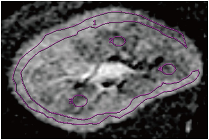
Fig. 1 ROI placement in transplanted kidney.ROI (1) as large as possible (about 500 pixels) is placed in renal cortex and several elliptical ROIs (2–4) with about 25 pixels each are placed in renal medulla. ROI = region of interest
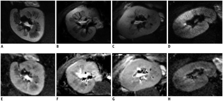
Fig. 2 DWI images and ADC maps of transplanted kidneys in different groups.DWI images (A–D) with b value of 10 s/mm2 with high resolution (1 × 1 mm2 for axial images) and ADC (E–H) maps of kidneys in 4 groups. Images from healthy volunteer (A, E) with eGFR = 102.1 mL/min/1.73 m2 and patient (B, F) in Group 1 with eGFR = 97.1 mL/min/1.73 m2 show marked corticomedullary differences. Images (C, G) from patient (eGFR = 54.59 mL/min/1.73 m2) in Group 2 show corticomedullary difference to lesser extent. Images (D, H) from patient (eGFR = 17.00 mL/min/1.73 m2) in Group 3 show no marked corticomedullary difference. ADC = apparent diffusion coefficient, DWI = diffusion-weighted imaging, eGFR = estimated glomerular filtration rate
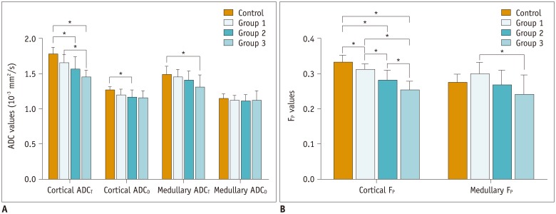
Fig. 3 Diffusion parameters in different groups.A. Cortical ADCT differs significantly among eGFR levels (p < 0.001). ADCT in Group 1 is significantly higher than Group 3 (p < 0.05). B. Cortical FP differs significantly among eGFR levels (p < 0.001), cortical FP values between each two groups show significant differences (p < 0.05). Medullary FP is significantly higher in Group 1 compared to Group 3 (p < 0.05). ADCD = perfusion-free apparent diffusion coefficient, ADCT = total apparent diffusion coefficient, FP = perfusion fraction
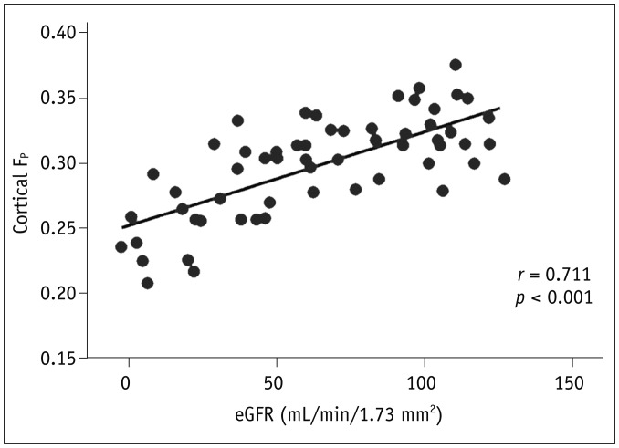
Fig. 4 Correlation analysis results between cortical FP and eGFR.FP values in cortex correlate positively with eGFR (r = 0.711, p < 0.001).
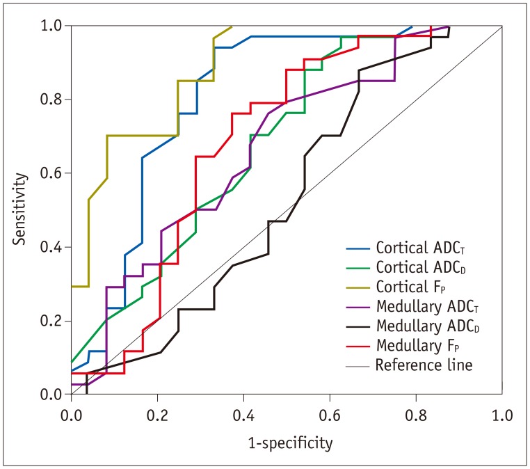
Fig. 5 Receiver operating characteristic curves of diffusion parameters.Using diffusion parameters as predictors for normal and mildly decreased renal function (eGFR ≥ 60 ml/min/1.73 m2), cortical FP shows best performance with area under curve of 0.890.
Cited by 1 articles
-
Multiparametric Functional Magnetic Resonance Imaging for Evaluating Renal Allograft Injury
Yuan Meng Yu, Qian Qian Ni, Zhen Jane Wang, Meng Lin Chen, Long Jiang Zhang
Korean J Radiol. 2019;20(6):894-908. doi: 10.3348/kjr.2018.0540.
Reference
-
1. Thoeny HC, De Keyzer F. Diffusion-weighted MR imaging of native and transplanted kidneys. Radiology. 2011; 259:25–38. PMID: 21436095.
Article2. Inci MF, Ozkan F, See TC, Tatli S. Renal transplant complications: diagnostic and therapeutic role of radiology. Can Assoc Radiol J. 2014; 65:242–252. PMID: 24325923.
Article3. Aktaş A. Transplanted kidney function evaluation. Semin Nucl Med. 2014; 44:129–145. PMID: 24484750.
Article4. Singh AK, Sahani DV. Imaging of the renal donor and transplant recipient. Radiol Clin North Am. 2008; 46:79–93. viPMID: 18328881.
Article5. Sharfuddin A. Renal relevant radiology: imaging in kidney transplantation. Clin J Am Soc Nephrol. 2014; 9:416–429. PMID: 24202132.
Article6. Sharfuddin A. Imaging evaluation of kidney transplant recipients. Semin Nephrol. 2011; 31:259–271. PMID: 21784275.
Article7. Thoeny HC, Zumstein D, Simon-Zoula S, Eisenberger U, De Keyzer F, Hofmann L, et al. Functional evaluation of transplanted kidneys with diffusion-weighted and BOLD MR imaging: initial experience. Radiology. 2006; 241:812–821. PMID: 17114628.
Article8. Le Bihan D, Breton E, Lallemand D, Aubin ML, Vignaud J, Laval-Jeantet M. Separation of diffusion and perfusion in intravoxel incoherent motion MR imaging. Radiology. 1988; 168:497–505. PMID: 3393671.
Article9. Abou-El-Ghar ME, El-Diasty TA, El-Assmy AM, Refaie HF, Refaie AF, Ghoneim MA. Role of diffusion-weighted MRI in diagnosis of acute renal allograft dysfunction: a prospective preliminary study. Br J Radiol. 2012; 85:e206–e211. PMID: 22215880.
Article10. Eisenberger U, Thoeny HC, Binser T, Gugger M, Frey FJ, Boesch C, et al. Evaluation of renal allograft function early after transplantation with diffusion-weighted MR imaging. Eur Radiol. 2010; 20:1374–1383. PMID: 20013274.
Article11. Eisenberger U, Binser T, Thoeny HC, Boesch C, Frey FJ, Vermathen P. Living renal allograft transplantation: diffusion-weighted MR imaging in longitudinal follow-up of the donated and the remaining kidney. Radiology. 2014; 270:800–808. PMID: 24475796.
Article12. Vermathen P, Binser T, Boesch C, Eisenberger U, Thoeny HC. Three-year follow-up of human transplanted kidneys by diffusion-weighted MRI and blood oxygenation level-dependent imaging. J Magn Reson Imaging. 2012; 35:1133–1138. PMID: 22180302.
Article13. Saritas EU, Cunningham CH, Lee JH, Han ET, Nishimura DG. DWI of the spinal cord with reduced FOV single-shot EPI. Magn Reson Med. 2008; 60:468–473. PMID: 18666126.
Article14. Levey AS, Stevens LA, Schmid CH, Zhang YL, Castro AF 3rd, Feldman HI, et al. A new equation to estimate glomerular filtration rate. Ann Intern Med. 2009; 150:604–612. PMID: 19414839.
Article15. Thoeny HC, De Keyzer F, Oyen RH, Peeters RR. Diffusion-weighted MR imaging of kidneys in healthy volunteers and patients with parenchymal diseases: initial experience. Radiology. 2005; 235:911–917. PMID: 15845792.
Article16. Yang D, Ye Q, Williams DS, Hitchens TK, Ho C. Normal and transplanted rat kidneys: diffusion MR imaging at 7 T. Radiology. 2004; 231:702–709. PMID: 15163810.
Article17. Dyvorne HA, Galea N, Nevers T, Fiel MI, Carpenter D, Wong E, et al. Diffusion-weighted imaging of the liver with multiple b values: effect of diffusion gradient polarity and breathing acquisition on image quality and intravoxel incoherent motion parameters--a pilot study. Radiology. 2013; 266:920–929. PMID: 23220895.18. Kang KM, Lee JM, Yoon JH, Kiefer B, Han JK, Choi BI. Intravoxel incoherent motion diffusion-weighted MR imaging for characterization of focal pancreatic lesions. Radiology. 2014; 270:444–453. PMID: 24126370.
Article19. Sigmund EE, Vivier PH, Sui D, Lamparello NA, Tantillo K, Mikheev A, et al. Intravoxel incoherent motion and diffusion-tensor imaging in renal tissue under hydration and furosemide flow challenges. Radiology. 2012; 263:758–769. PMID: 22523327.
Article20. Joo I, Lee JM, Han JK, Choi BI. Intravoxel incoherent motion diffusion-weighted MR imaging for monitoring the therapeutic efficacy of the vascular disrupting agent CKD-516 in rabbit VX2 liver tumors. Radiology. 2014; 272:417–426. PMID: 24697148.
Article21. Heusch P, Wittsack HJ, Heusner T, Buchbender C, Quang MN, Martirosian P, et al. Correlation of biexponential diffusion parameters with arterial spin-labeling perfusion MRI: results in transplanted kidneys. Invest Radiol. 2013; 48:140–144. PMID: 23249648.22. Le Bihan D, Turner R. The capillary network: a link between IVIM and classical perfusion. Magn Reson Med. 1992; 27:171–178. PMID: 1435202.23. Toya R, Naganawa S, Kawai H, Ikeda M. Correlation between estimated glomerular filtration rate (eGFR) and apparent diffusion coefficient (ADC) values of the kidneys. Magn Reson Med Sci. 2010; 9:59–64. PMID: 20585195.
Article24. Blondin D, Lanzman RS, Mathys C, Grotemeyer D, Voiculescu A, Sandmann W, et al. [Functional MRI of transplanted kidneys using diffusion-weighted imaging]. Rofo. 2009; 181:1162–1167. PMID: 19582653.25. Cosio FG, Grande JP, Larson TS, Gloor JM, Velosa JA, Textor SC, et al. Kidney allograft fibrosis and atrophy early after living donor transplantation. Am J Transplant. 2005; 5:1130–1136. PMID: 15816896.
Article
- Full Text Links
-
- Actions
-
Cited
- CITED
-
- Close
- Share
-
- Similar articles
-
- High field strength magnetic resonance imaging of brain lesion
- Single-Shot Echo-Planar Diffusion-Weighted MR Imaging at 3T and 1.5T for Differentiation of Benign Vertebral Fracture Edema and Tumor Infiltration
- Assessment of Diffusion-Weighted Imaging-FLAIR Mismatch: Comparison between Conventional FLAIR versus Shorter-Repetition-Time FLAIR at 3T
- Small Focal Hepatic Lesions <=1 cm: Their Detection and Characterization with Performing Diffusion-Weighted Sensitivity-Encoding versus SPIO-Enhanced 3T MR Imaging
- Basics for Pediatric Brain Tumor Imaging: Techniques and Protocol Recommendations
