J Pathol Transl Med.
2018 Jan;52(1):56-60. 10.4132/jptm.2017.04.08.
Colloid Carcinoma of the Uterine Cervix and Its Immunohistochemical Analysis: A Case Report
- Affiliations
-
- 1Department of Pathology, Zeynep Kamil Maternity and Pediatric Research and Training Hospital, Istanbul, Turkey.
- 2Department of Obstetrics and Gynecology, Zeynep Kamil Maternity and Pediatric Research and Training Hospital, Istanbul, Turkey. pataraa96@gmail.com
- 3Department of Pathology, Fatih Sultan Mehmet Research and Training Hospital, Istanbul, Turkey.
- KMID: 2403259
- DOI: http://doi.org/10.4132/jptm.2017.04.08
Abstract
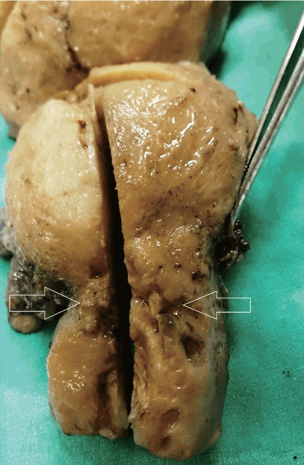
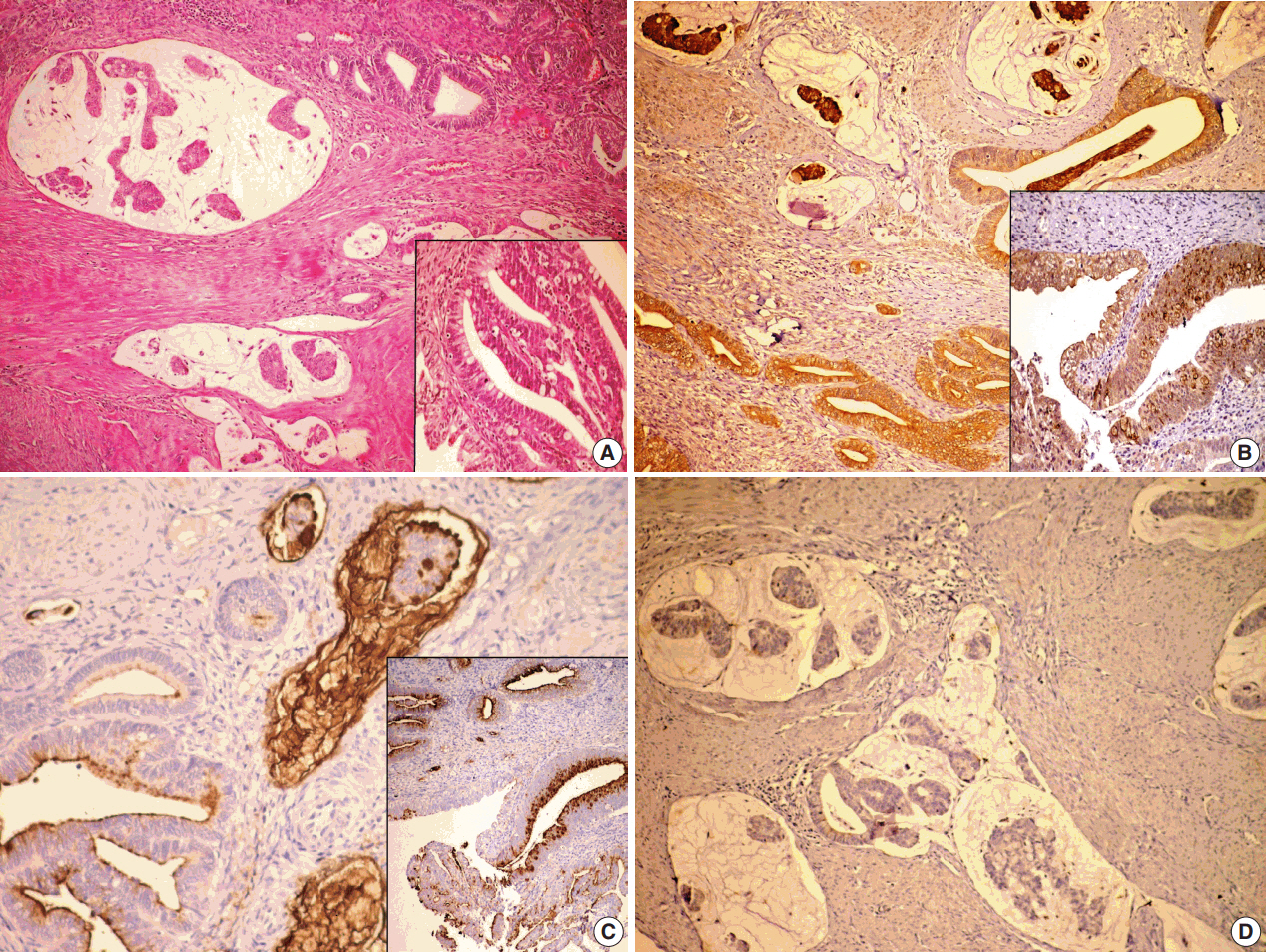
- Colloid carcinoma, which is a very rare tumor of the uterine cervix, is composed of an excessive amount of mucus and a relative paucity of tumoral glandular cells within them. Herein, we report a rare case of colloid carcinoma of the cervix with adenocarcinoma in situ (AIS), intestinal and usual types, and endocervical adenocarcinoma (usual type) components. We also discuss the morphological and immunohistochemical characteristics of this tumor. A 51-year-old woman was referred to our outpatient clinic with the symptom of genital bleeding lasting for 5 months. She had a cervix surrounded by an irregular tumor with a diameter of 5 cm. The colloid carcinoma cells were positive for MUC2, MUC5AC, and cytokeratin (CK) 7, focal positive for CDX2, and negative for MUC6 and CK20. Also, the intestinal type AIS showed a similar staining pattern. Colloid carcinoma cells producing mucin showed an intestinal phenotype and AIS. The intestinal type can be considered as a precursor lesion of colloid carcinoma.
Keyword
MeSH Terms
Figure
Reference
-
1. Shintaku M, Kushima R, Abiko K. Colloid carcinoma of the intestinal type in the uterine cervix: mucin immunohistochemistry. Pathol Int. 2010; 60:119–24.
Article2. Ishida M, Iwai M, Kagotani A, Yoshida K, Okabe H. Colloid carcinoma of the uterine cervix: a case report with respect to immunohistochemical analyses. Int J Gynecol Pathol. 2014; 33:248–52.3. Walker AN, Mills SE. Unusual variants of uterine cervical carcinoma. Pathol Annu. 1987; 22 Pt 1:277–310.4. Hurt WG, Silverberg SG, Frable WJ, Belgrad R, Crooks LD. Adenocarcinoma of the cervix: histopathologic and clinical features. Am J Obstet Gynecol. 1977; 129:304–15.
Article5. Kurman RJ, Carcangiu ML, Herrington CS, Young RH. WHO classification of tumors of female reproductive organs. Lyon: International Agency for Research on Cancer;2014.6. Young RH, Clement PB. Endocervical adenocarcinoma and its variants: their morphology and differential diagnosis. Histopathology. 2002; 41:185–207.
Article7. Riethdorf L, O’Connell JT, Riethdorf S, Cviko A, Crum CP. Differential expression of MUC2 and MUC5AC in benign and malignant glandular lesions of the cervix uteri. Virchows Arch. 2000; 437:365–71.
Article8. Baker AC, Eltoum I, Curry RO, et al. Mucinous expression in benign and neoplastic glandular lesions of the uterine cervix. Arch Pathol Lab Med. 2006; 130:1510–5.
Article9. Bu XD, Li N, Tian XQ, et al. Altered expression of MUC2 and MUC5AC in progression of colorectal carcinoma. World J Gastroenterol. 2010; 16:4089–94.
Article10. McCluggage WG, Shah R, Connolly LE, McBride HA. Intestinaltype cervical adenocarcinoma in situ and adenocarcinoma exhibit a partial enteric immunophenotype with consistent expression of CDX2. Int J Gynecol Pathol. 2008; 27:92–100.
Article11. Sullivan LM, Smolkin ME, Frierson HF Jr, Galgano MT. Comprehensive evaluation of CDX2 in invasive cervical adenocarcinomas: immunopositivity in the absence of overt colorectal morphology. Am J Surg Pathol. 2008; 32:1608–12.
- Full Text Links
- Actions
-
Cited
- CITED
-
- Close
- Share
- Similar articles
-
- Adenoid Basal Carcinoma Associated with Invasive Squamous Cell Carcinoma of Uterine Cervix: A case report
- Adenoid Basal Carcinoma Associated with Invasive Squamous Cell Carcinoma of Uterine Cervix: A case report
- Verrucous Carcinoma of Uterine Cervix: A case report
- A Case of Lymphoepithelioma - Like Carcinoma of Uterine Cervix
- A Case of Adenoid Basal Carcinoma of the Uterine Cervix


