Semi-Functional Quantitative Flow Cytometry Assay for Lymphocytic Choriomeningitis Virus Titration
- Affiliations
-
- 1Department of Biochemistry, College of Life Science & Biotechnology, Yonsei University Seoul 03722, Korea. sjha@yonsei.ac.kr
- KMID: 2400636
- DOI: http://doi.org/10.4110/in.2017.17.5.307
Abstract
- Quantitative PCR and plaque assay are powerful virological techniques used to measure the load of defective or infectious virus in mouse and human. However, these methods display limitations such as cross contamination and long run-time. Here, we describe a novel technique termed as semi-functional quantitative flow cytometry (SFQF) for the accurate estimation of the quantity of infectious lymphocytic choriomeningitis virus (LCMV). LCMV titration method using flow cytometry was previously developed but has technical shortcomings, owing to the less optimized parameters such as cell overgrowth, plate scale, and detection threshold. Therefore, we first established optimized conditions for SFQF assay using LCMV nucleoprotein (NP)-specific antibody to evaluate the threshold of the virus detection range in the plaque assay. We subsequently demonstrated that the optimization of the method increased the sensitivity of virus detection. We revealed several new advantages of SFQF assay, which overcomes some of the previously contentious points, and established an upgraded version of the previously reported flow cytometric titration assay. This method extends the detection scale to the level of single cell, allowing extension of its application for in vivo detection of infected cells and their phenotypic analysis. Thus, SFQF assay may serve as an alternative analytical tool for ensuring the reliability of LCMV titration and can be used with other types of viruses using target-specific antibodies.
Keyword
MeSH Terms
Figure
-
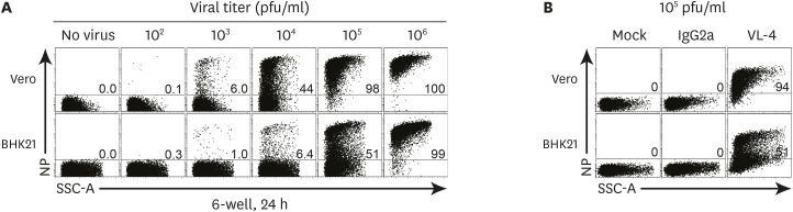
Figure 1 Vero cells were more susceptible than BHK21 cells to LCMV ARM infection. A total of 3.0×10⁵ Vero and BHK21 cells were plated in a 6-well plate with 3 ml of DMEM complete medium for 15 h before use. After the removal of the culture medium, cells were treated with 200 µl of serial dilutions (106 to 102 pfu/ml) of LCMV ARM for 1 h. A total of 2 ml of the medium was dispensed and cells were stained with VL-4 after 24 h of incubation, followed by flow cytometry analysis. (A) NP expression in Vero and BHK21 cells infected with serial dilutions of LCMV ARM. (B) The expression of isotype control of VL-4 antibody (IgG2a) in Vero and BHK21 cells infected with LCMV ARM (105 pfu/ml). The number in the plot indicates the percentage of NP+ cell population in Vero and BHK21 cells. Data are representative of 5 independent experiments.SSC-A, side scatter area.
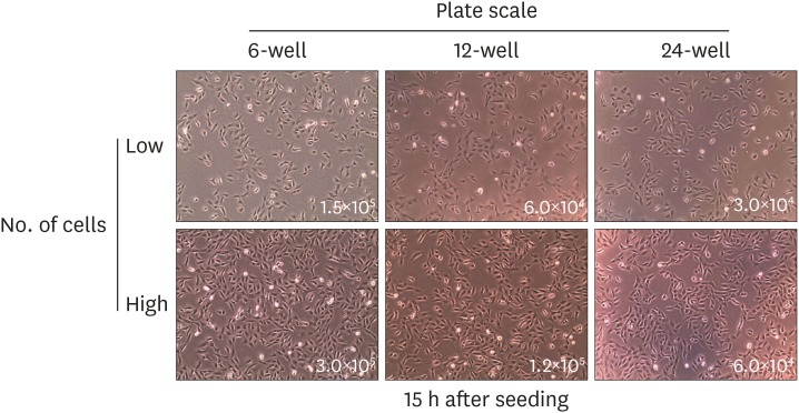
Figure 2 Initial cell seeding number was set to high and low for each plate scale. Cell numbers for each scale were calculated in proportion to the size of 6-, 12-, and 24-well plate. Cells were seeded and incubated for 15 h prior to infection. The number in the image visualized by an optical microscope indicates the cell count of seeded cells as high (top) and low (bottom) confluence in each plate. Data are representative of 2 independent experiments.
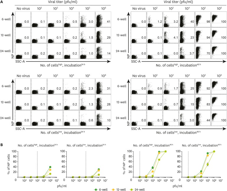
Figure 3 Validation of conditions such as plate scale, cell seeding number, and incubation time to establish an optimal method for SFQF assay. Vero cells were plated on 6-, 12-, and 24-well plates at high and low seeding numbers (as previously determined) and cultured for 15 h. The medium was removed and cells were treated with 200 µl of the serial dilution of LCMV ARM stock (106 pfu/ml) at each incubation time, followed by flow cytometry analysis. (A) The incubation of 48 h in a 6-well plate seeded with a high number of cells was optimal for LCMV detection. Numbers in representative plots indicate percentages of NP+ cells at indicated conditions such as cell seeding number and incubation time (left top, high and 24 h; left bottom, low and 24 h; right top, high and 48 h; right bottom, low and 48 h). (B) Line graphs summarize the representative data of Fig. 3A obtained from each condition. Data are representatives of 2 independent experiments.SSC-A, side scatter area.
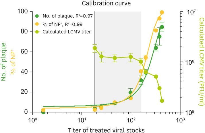
Figure 4 Plaque numbers and NP+ cell frequencies were correlated to obtain a standard curve. Vero cells were plated as per the conditions in Fig. 3 and infected with 200 µl of 2-fold dilution of LCMV ARM (106 pfu/ml) in the amount same as that in the plaque assay. After 48 h of incubation, the frequency of NP+ cells was analyzed by flow cytometry. All of the serial dilutions were analyzed by plaque assay to verify the threshold of the virus detection. Calibration curve of NP+ cell frequency (yellow circle) and plaque number (green circle). Calculation of plaque assay for each serial dilution (yellow green circle). Left Y-axis, right Y-axis, and X-axis indicate plaque number with NP+ cell frequency, calculated LCMV titer of each serial dilution, and titer of each treated serial dilution, respectively. R2 values were obtained using non-linear curve fit analysis. Data are representative of 3 independent experiments.

Figure 5 Validation of LCMV titration in tissues ex vivo. C57BL/6 mice were intravenously infected with LCMV CL13 (2×10⁶ pfu/ml). Spleens and serum were harvested from mice 10 days after infection. Homogenized spleens and bled serum from 10−4 to 10−2 dilution of original stocks were incubated with Vero cells and SFQF and plaque assays were manually performed. (A) Detection of NP+ proportion in serum and spleens of mice infected with LCMV CL13. Numbers in plots indicate the percentage of NP+ population. (B) Bar graphs summarize the titer calculated from the plaque assay (green bar) and frequency of NP+ cells measured by SFQF assay (yellow bar). n=3 mice per group in each experiment.
Reference
-
1. Zhou X, Ramachandran S, Mann M, Popkin DL. Role of lymphocytic choriomeningitis virus (LCMV) in understanding viral immunology: past, present and future. Viruses. 2012; 4:2650–2669. PMID: 23202498.
Article2. Salvato MS, Shimomaye EM. The completed sequence of lymphocytic choriomeningitis virus reveals a unique RNA structure and a gene for a zinc finger protein. Virology. 1989; 173:1–10. PMID: 2510401.
Article3. Pinschewer DD, Perez M, de la Torre JC. Role of the virus nucleoprotein in the regulation of lymphocytic choriomeningitis virus transcription and RNA replication. J Virol. 2003; 77:3882–3887. PMID: 12610166.
Article4. Darbre S, Johnson S, Kallert S, Lambert PH, Siegrist CA, Pinschewer DD. The nucleoprotein is required for lymphocytic choriomeningitis virus-based vaccine vector immunogenicity. J Virol. 2015; 89:11734–11738. PMID: 26355095.
Article5. Marcus PI, Carver DH. Hemadsorption-negative plaque test: new assay for rubella virus revealing a unique interference. Science. 1965; 149:983–986. PMID: 4283964.
Article6. Cooper PD. The plaque assay of animal viruses. Adv Virus Res. 1961; 8:319–378. PMID: 13881155.
Article7. Sedwick WD, Wiktor TJ. Reproducible plaquing system for rabies, lymphocytic choriomeningitis,k and other ribonucleic acid viruses in BHK-21-13S agarose suspensions. J Virol. 1967; 1:1224–1226. PMID: 4987176.8. Wainwright S, Mims CA. Plaque assay for lymphocytic choriomeningitis virus based on hemadsorption interference. J Virol. 1967; 1:1091–1092. PMID: 4987174.
Article9. McCausland MM, Crotty S. Quantitative PCR technique for detecting lymphocytic choriomeningitis virus in vivo. J Virol Methods. 2008; 147:167–176. PMID: 17920702.10. Cordey S, Sahli R, Moraz ML, Estrade C, Morandi L, Cherpillod P, Charrel RN, Kunz S, Kaiser L. Analytical validation of a lymphocytic choriomeningitis virus real-time RT-PCR assay. J Virol Methods. 2011; 177:118–122. PMID: 21763351.
Article11. Welsh RM, Pfau CJ. Determinants of lymphocytic choriomeningitis interference. J Gen Virol. 1972; 14:177–187. PMID: 4622135.
Article12. Welsh RM Jr, Buchmeier MJ. Protein analysis of defective interfering lymphocytic choriomeningitis virus and persistently infected cells. Virology. 1979; 96:503–515. PMID: 380148.
Article13. Welsh RM, Oldstone MB. Inhibition of immunologic injury of cultured cells infected with lymphocytic choriomeningitis virus: role of defective interfering virus in regulating viral antigenic expression. J Exp Med. 1977; 145:1449–1468. PMID: 301173.
Article14. Sheĭnbergas MM, Vorob’eva ZN. Titration of antibodies to lymphocytic choriomeningitis virus by the method of indirect immunofluorescence. Vopr Virusol. 1975; 357–360. PMID: 1099805.15. Battegay M, Cooper S, Althage A, Bänziger J, Hengartner H, Zinkernagel RM. Quantification of lymphocytic choriomeningitis virus with an immunological focus assay in 24- or 96-well plates. J Virol Methods. 1991; 33:191–198. PMID: 1939506.
Article16. Battegay M. Quantification of lymphocytic choriomeningitis virus with an immunological focus assay in 24 well plates. ALTEX. 1993; 10:6–14. PMID: 11178349.17. Korns Johnson D, Homann D. Accelerated and improved quantification of lymphocytic choriomeningitis virus (LCMV) titers by flow cytometry. PLoS One. 2012; 7:e37337. PMID: 22615984.
Article18. Dutko FJ, Oldstone MB. Genomic and biological variation among commonly used lymphocytic choriomeningitis virus strains. J Gen Virol. 1983; 64:1689–1698. PMID: 6875516.
Article19. Ahmed R, Salmi A, Butler LD, Chiller JM, Oldstone MB. Selection of genetic variants of lymphocytic choriomeningitis virus in spleens of persistently infected mice. Role in suppression of cytotoxic T lymphocyte response and viral persistence. J Exp Med. 1984; 160:521–540. PMID: 6332167.
Article20. Cao W, Henry MD, Borrow P, Yamada H, Elder JH, Ravkov EV, Nichol ST, Compans RW, Campbell KP, Oldstone MB. Identification of alpha-dystroglycan as a receptor for lymphocytic choriomeningitis virus and Lassa fever virus. Science. 1998; 282:2079–2081. PMID: 9851928.21. Welsh RM, Seedhom MO. Lymphocytic choriomeningitis virus (LCMV): propagation, quantitation, and storage. Curr Protoc Microbiol. 2008; Chapter 15:Unit 15A.1.
Article22. Bocharov G, Ludewig B, Bertoletti A, Klenerman P, Junt T, Krebs P, Luzyanina T, Fraser C, Anderson RM. Underwhelming the immune response: effect of slow virus growth on CD8+-T-lymphocyte responses. J Virol. 2004; 78:2247–2254. PMID: 14963121.23. Ammerman NC, Beier-Sexton M, Azad AF. Growth and maintenance of Vero cell lines. Curr Protoc Microbiol . Appendix. 2008; Appendix 4:Appendix 4E.
- Full Text Links
-
- Actions
-
Cited
- CITED
-
- Close
- Share
-
- Similar articles
-
- Comparative Study of the Standard Plaque Assay with Solid-overlay and Immunofocus Assay for Varicella-zoster Virus Titration
- Extrinsic Acquisition of CD80 by Antigen-Specific CD8⺠T Cells Regulates Their Recall Immune Responses to Acute Viral Infection
- The Multifaceted Roles of NK Cells in the Context of Murine Cytomegalovirus and Lymphocytic Choriomeningitis Virus Infections
- Re-defining T-Cell Exhaustion: Subset, Function, and Regulation
- Metabolic Reprogramming by the Excessive AMPK Activation Exacerbates Antigen-Specific Memory CD8⺠T Cell Differentiation after Acute Lymphocytic Choriomeningitis Virus Infection
