J Cardiovasc Ultrasound.
2017 Dec;25(4):140-141. 10.4250/jcu.2017.25.4.140.
Congenital Left Atrial Bands with Atrial Fibrillation
- Affiliations
-
- 1Cardiovascular Center, Korea University Guro Hospital, Seoul, Korea. koolup93@gmail.com
- KMID: 2399416
- DOI: http://doi.org/10.4250/jcu.2017.25.4.140
Abstract
- No abstract available.
MeSH Terms
Figure
-
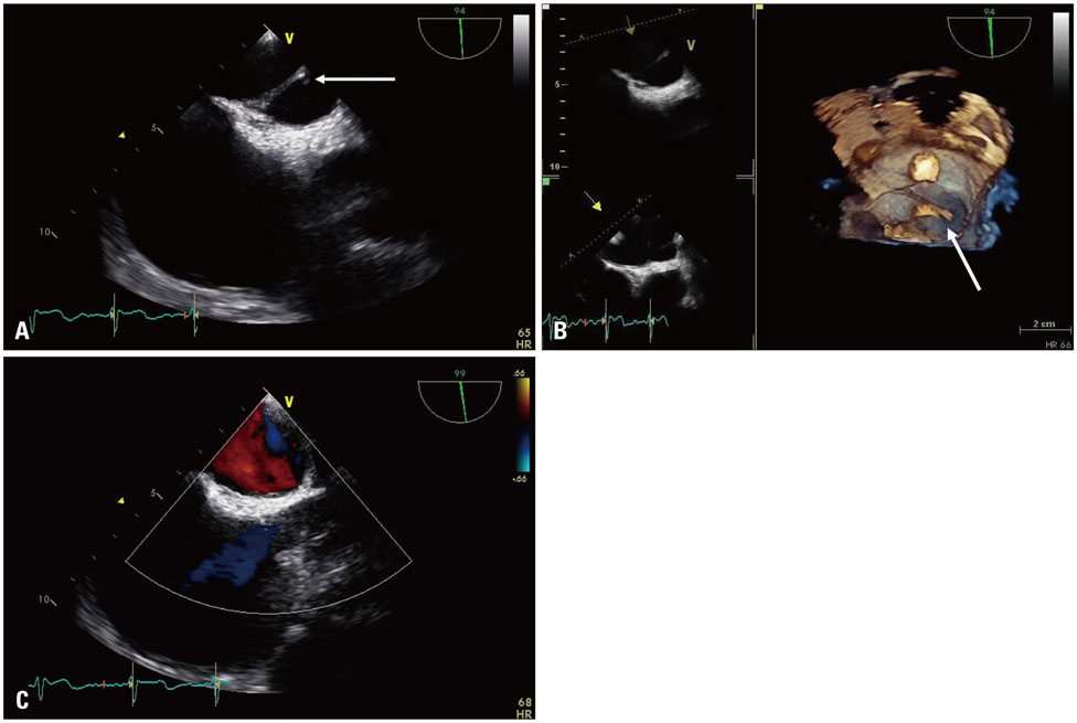
Fig. 1 TEE showed non-mobile linear structures in the left atrium. A. Two-dimensional TEE (white arrow). B. 3D TEE (white arrow) and 3D view direction (yellow arrow). C. There was no flow limitation or acceleration on Doppler study. TEE: transesophageal echocardiography, 3D: three-dimensional.
Reference
-
1. Yamashita T, Ohkawa S, Imai T, Ide H, Watanabe C, Ueda K. Prevalence and clinical significance of anomalous muscular band in the left atrium. Am J Cardiovasc Pathol. 1993; 4:286–293.2. Baran T, Küçükoğlu MS, Okçün B, Cetin G, Hatemi AC, Uner S. A rare cause of mitral insufficiency: left atrial anomalous band. Echocardiography. 2003; 20:83–85.3. McNamara WL, Baker LA, Costich K. Asymptomatic congenital anomaly of the heart; congenital muscular cord bridging walls of auricle above center of mitral valve. Am Heart J. 1947; 34:288–290.4. Ozer O, Sari I, Davutoglu V, Yigiter R, Akkoyun C. Cryptogenic stroke in two cases with left atrial band: coincidence or cause. Eur J Echocardiogr. 2009; 10:360–361.5. Uetake S, Miyauchi Y, Hayashi M, Shimizu W. Electrophysiological characteristics of a left atrial anomalous muscular band in a case with paroxysmal atrial fibrillation. HeartRhythm Case Rep. 2015; 1:78–81.
- Full Text Links
-
- Actions
-
Cited
- CITED
-
- Close
- Share
-
- Similar articles
-
- Relation between Atrial Fibrillation and Echocardiographic Size of Left Atrium
- Persistent Atrial Fibrillation Related to a Congenital Pericardial Defect and Left Atrial Appendage Herniation
- The Influence of Electrical Cardioversion for Atrial Fibrillation on Left Atrial Appendage Function: A Transesophageal Echocardiography Study
- Atrial Fibrillation in a Patient with Left Ventricular Hypertrophy after Induction of General Anesthesia: A case report
- Pathophysiology and Diagnosis in Atrial Fibrillation
