Ghost cell odontogenic carcinoma on right mandible and its respective surgical reconstruction: a case report
- Affiliations
-
- 1Department of Oral and Maxillofacial Surgery, School of Dentistry, Seoul National University, Seoul, Korea. myoungh@snu.ac.kr
- 2Dental Research Institute, Seoul National University, Seoul, Korea.
- KMID: 2398997
- DOI: http://doi.org/10.5125/jkaoms.2017.43.6.415
Abstract
- Calcifying cystic odontogenic tumor (CCOT) is defined as an odontogenic cyst-like benign neoplasm that characteristically contains several ghost cells, ameloblastoma-like epithelium, and occasional calcification. Ghost cell odontogenic carcinoma (GCOC), a malignant form of CCOT, is an exceptionally rare malignant tumor. In this report, we present a case of a 53-year-old man whose chief complaint was a solitary mass on the right mandible area. The mass was completely removed through an extraoral surgical approach and reconstructive surgery was performed in two phases.
Keyword
Figure
-
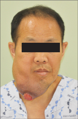
Fig. 1 Preoperative extraoral clinical photo.

Fig. 2 Preoperative intraoral clinical photo.
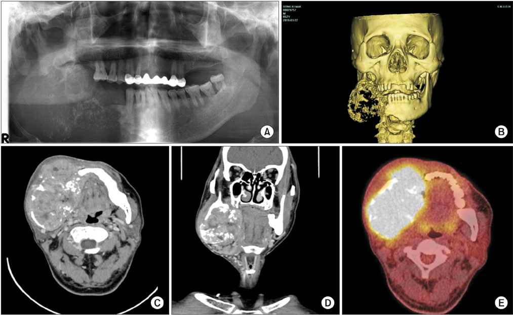
Fig. 3 Clinical and radiologic findings. A. Panoramic view. B. Preoperative three-dimensional computed tomography (CT) view. C. Preoperative enhanced CT view (axial cut). D. Preoperative enhanced CT view (coronal cut). E. Preoperative positron emission tomography-CT.

Fig. 4 A. Post mass resection. B. Main mass.
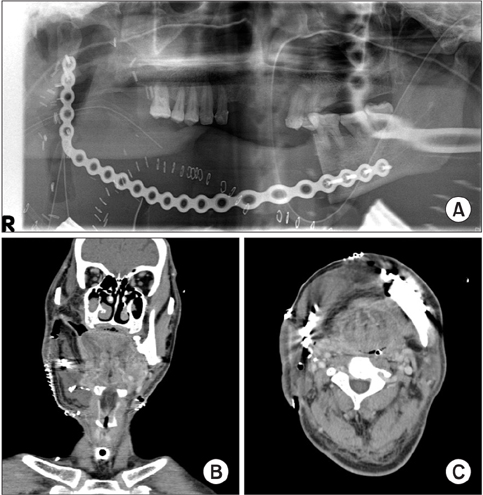
Fig. 5 Radiographic findings after first surgery. A. Panoramic view after first surgery. B. Enhanced computed tomography (CT) view (axial view) after first surgery. C. Enhanced CT view (coronal view) after first surgery.
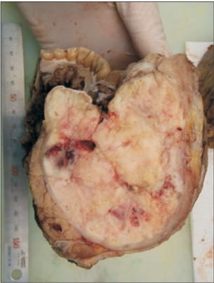
Fig. 6 Main mass.
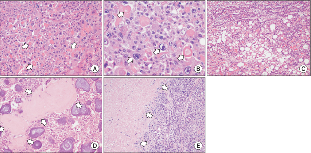
Fig. 7 Histologic findings. A. Malignant epithelial cells with ghost cells (arrows; H&E staining, ×200). B. Atypical cells with prominent pleomorphism (arrows; H&E staining, ×400). C. Long odontogenic epithelial strands with focal clear cell clusters (H&E staining, ×100). D. Dysplastic dentin with concentric calcifications (arrows; H&E staining, ×200). E. Focal necrosis (arrows; H&E staining, ×100).

Fig. 8 Histologic findings. A. Immunohistochemical staining (Ki-67). B. Immunohistochemical staining (pan-cytokeratin).
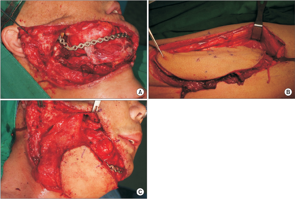
Fig. 9 Second reconstruction surgery. A. Previous flap removal. B. Flap harvesting. C. Flap setting.
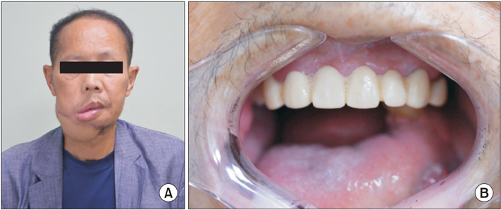
Fig. 10 Six month postoperative follow-up. A. Extraoral clinical photo. B. Intraoral clinical photo.
Reference
-
1. Gorlin RJ, Pindborg JJ, Odont , Clausen FP, Vickers RA. The calcifying odontogenic cyst: a possible analogue of the cutaneous calcifying epithelioma of Malherbe. An analysis of fifteen cases. Oral Surg Oral Med Oral Pathol. 1962; 15:1235–1243.
Article2. Nazaretian SP, Schenberg ME, Simpson I, Slootweg PJ. Ghost cell odontogenic carcinoma. Int J Oral Maxillofac Surg. 2007; 36:455–458.
Article3. Tarakji B, Ashok N, Alzoghaibi I, Altamimi MA, Azzeghaiby SN, Baroudi K, et al. Malignant transformation of calcifying cystic odontogenic tumour: a review of literature. Contemp Oncol (Pozn). 2015; 19:184–186.4. Martos-Fernández M, Alberola-Ferranti M, Hueto-Madrid JA, Bescós-Atín C. Ghost cell odontogenic carcinoma: a rare case report and review of literature. J Clin Exp Dent. 2014; 6:e602–e606.
Article5. Motosugi U, Ogawa I, Yoda T, Abe T, Sugasawa M, Murata S, et al. Ghost cell odontogenic carcinoma arising in calcifying odontogenic cyst. Ann Diagn Pathol. 2009; 13:394–397.
Article6. Lee SK, Kim YS. Current concepts and occurrence of epithelial odontogenic tumors: II. calcifying epithelial odontogenic tumor versus ghost cell odontogenic tumors derived from calcifying odontogenic cyst. Korean J Pathol. 2014; 48:175–187.
Article7. Cheng Y, Long X, Li X, Bian Z, Chen X, Yang X. Clinical and radiological features of odontogenic ghost cell carcinoma: review of the literature and report of four new cases. Dentomaxillofac Radiol. 2004; 33:152–157.
Article8. Arashiyama T, Kodama Y, Kobayashi T, Hoshina H, Takagi R, Hayashi T, et al. Ghost cell odontogenic carcinoma arising in the background of a benign calcifying cystic odontogenic tumor of the mandible. Oral Surg Oral Med Oral Pathol Oral Radiol. 2012; 114:e35–e40.
Article9. Bianchi B, Copelli C, Ferrari S, Ferri A, Sesenna E. Free flaps: outcomes and complications in head and neck reconstructions. J Craniomaxillofac Surg. 2009; 37:438–442.
Article10. Pohlenz P, Klatt J, Schön G, Blessmann M, Li L, Schmelzle R. Microvascular free flaps in head and neck surgery: complications and outcome of 1000 flaps. Int J Oral Maxillofac Surg. 2012; 41:739–743.
Article11. Zhang C, Sun J, Zhu H, Xu L, Ji T, He Y, et al. Microsurgical free flap reconstructions of the head and neck region: Shanghai experience of 34 years and 4640 flaps. Int J Oral Maxillofac Surg. 2015; 44:675–684.
Article12. Ozkan O, Ozkan O, Coskunfirat OK, Hadimioğlu N. Reconstruction of large palatal defects using the free anterolateral thigh flap. Ann Plast Surg. 2011; 66:618–622.
Article13. Leclère FM, Bosc R, Temam S, Leymarie N, Mirghani H, Sarfati B, et al. Reconstruction of large mandibulofacial defects with the composed double skin paddle fibula free flap: a review of 32 procedures. Laryngoscope. 2014; 124:1336–1343.
Article14. Myers LL, Ahn C. Does increased free flap size in the head and neck region impact clinical outcome? J Oral Maxillofac Surg. 2014; 72:1832–1840.
Article15. Cooley BC, Hanel DP, Anderson RB, Foster MD, Gould JS. The influence of diabetes on free flap transfer: I. Flap survival and microvascular healing. Ann Plast Surg. 1992; 29:58–64.16. Cooley BC, Hanel DP, Lan M, Li X, Gould JS. The influence of diabetes on free flap transfer: II. The effect of ischemia on flap survival. Ann Plast Surg. 1992; 29:65–69.17. Liu Z, Tian Z, Zhang C, Sun J, Zhang Z, He Y. Microvascular reconstruction in elderly oral cancer patients: does diabetes status have a predictive role in free flap complications? J Oral Maxillofac Surg. 2015; 73:357–369.
Article18. Rosado P, Cheng HT, Wu CM, Wei FC. Influence of diabetes mellitus on postoperative complications and failure in head and neck free flap reconstruction: a systematic review and meta-analysis. Head Neck. 2015; 37:615–618.
Article19. Genden EM, Rinaldo A, Suárez C, Wei WI, Bradley PJ, Ferlito A. Complications of free flap transfers for head and neck reconstruction following cancer resection. Oral Oncol. 2004; 40:979–984.
Article20. Bozikov K, Arnez ZM. Factors predicting free flap complications in head and neck reconstruction. J Plast Reconstr Aesthet Surg. 2006; 59:737–742.
Article
- Full Text Links
-
- Actions
-
Cited
- CITED
-
- Close
- Share
-
- Similar articles
-
- Odontogenic ghost cell carcinoma arising from odontogenic epithelial tumor in maxilla: A case report
- Dentinogenic Ghost Cell Tumor: A Case Report and Review of Literature
- Ghost Cell Odontogenic Carcinoma Arising from Calcifying Cystic Odontogenic Tumor: A Case Report
- Primary intraosseous carcinoma(PIOC) on mandible: Case Report
- Current Concepts and Occurrence of Epithelial Odontogenic Tumors: II. Calcifying Epithelial Odontogenic Tumor Versus Ghost Cell Odontogenic Tumors Derived from Calcifying Odontogenic Cyst
