Effectiveness of cholangioscopy using narrow band imaging for hepatobiliary malignancies
- Affiliations
-
- 1Department of Gastroenterology, Eulji University Hospital, Eulji University College of Medicine, Daejeon, Korea. jj98w@naver.com
- KMID: 2398958
- DOI: http://doi.org/10.4174/astr.2017.93.3.125
Abstract
- PURPOSE
Recently, cholangioscopy using narrow band imaging (NBI) has been used as a diagnostic modality for better visualization in hepatobiliary malignancies; however, there are few reports on it. Our aim is to evaluate the effectiveness of cholangioscopy using NBI in hepatobiliary malignancies.
METHODS
Between January 2007 and December 2016, 152 cholangioscopies using percutaneous approach were conducted in total 123 patients. Among these, 36 patients were suspicious of hepatobiliary malignancies. Thirteen patients with an ambiguous margin on endoscopic retrograde cholangiopancreatography (ERCP) or magnetic resonance cholangiopancreatography (MRCP), for whom NBI tipped the balance in diagnosis of lesion and decision of lesion extent by adding NBI, were involved in our study.
RESULTS
Underlying diseases were all malignant in 13 patients (11 bile duct cancers, 1 liver cancer, 1 pancreas cancer with common bile duct invasion). In 7 cases with papillary type tumor, minute superficial spreading tumor was detected by NBI more easily, and NBI provided a better visualization of tumor vessel and margin evaluation in 4 cases with infiltrative tumor. In 2 cases with mucin-hypersecreting tumor, NBI showed better penetration through the mucin and gave us a much clearer image. Nine patients ultimately underwent surgical resection. The margins predicted by NBI cholangioscopy were consistent with the pathological margins on the resected specimens.
CONCLUSION
In conclusion, cholangioscopy using NBI is very useful for evaluation of suspected hepatobiliary malignancies with an ambiguous margin on ERCP or MRCP. It can give us an accurate pathologic mapping, and this information seems to be essential before deciding on a treatment strategy.
Keyword
MeSH Terms
Figure
-

Fig. 1 Papillary type cancer. (A) Minute papillary mucosal changes (arrows) were suspected at this level of bile duct. But, the border of lesion was ambiguous. (B) With narrow band imaging, a prominent papillary lesion (arrowheads) was noted at the same level.
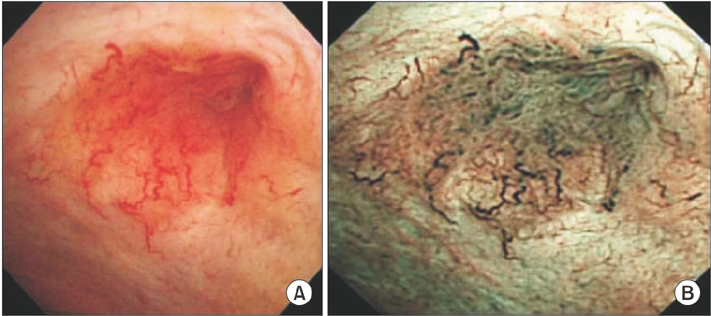
Fig. 2 Infiltrative type cancer. (A) Conventional white-light imaging showed a tapered, luminal narrowing of the common hepatic duct and neovascularization of its surface. (B) After narrow band imaging, more irregular mucosal surface, color change on the tumor vessel, and more vascular changes especially on the tumor margin were noted.

Fig. 3 Mucin-hypersecreting type tumor. (A) White-light imaging showed floating mucinous material, however, we could not identify any mucosal change because of the large amount of mucin. (B) On narrow band imaging, both mucin and the tumor mass were clearly visible.
Reference
-
1. Nimura Y, Kamiya J, Hayakawa N, Shionoya S. Cholangioscopic differentiation of biliary strictures and polyps. Endoscopy. 1989; 21:Suppl 1. 351–356.2. Neuhaus H. Cholangioscopy. Endoscopy. 1994; 26:120–125.3. Seo DW, Kim MH, Lee SK, Myung SJ, Kang GH, Ha HK, et al. Usefulness of cholangioscopy in patients with focal stricture of the intrahepatic duct unrelated to intrahepatic stones. Gastrointest Endosc. 1999; 49:204–209.4. Nimura Y, Kamiya J. Cholangioscopy. Endoscopy. 1998; 30:182–188.5. Kim YS, Myung SJ, Kim SY, Kim HJ, Kim JS, Park ET, et al. Biliary papillomatosis: clinical, cholangiographic and cholangioscopic findings. Endoscopy. 1998; 30:763–767.6. Jeng KS. Treatment of intrahepatic biliary stricture associated with hepatolithiasis. Hepatogastroenterology. 1997; 44:342–351.7. Sheen-Chen SM, Cheng YF, Chou FF, Lee TY. Ductal dilatation and stenting make routine hepatectomy unnecessary for left hepatolithiasis with intrahepatic biliary stricture. Surgery. 1995; 117:32–36.8. Jeng KS, Yang FS, Ohta I, Chiang HJ. Dilatation of intrahepatic biliary strictures in patients with hepatolithiasis. World J Surg. 1990; 14:587–592.9. ASGE Technology Committee. Song LM, Adler DG, Conway JD, Diehl DL, Farraye FA, et al. Narrow band imaging and multiband imaging. Gastrointest Endosc. 2008; 67:581–589.10. Itoi T, Sofuni A, Itokawa F, Tsuchiya T, Kurihara T, Ishii K, et al. Peroral cholangioscopic diagnosis of biliary-tract diseases by using narrow-band imaging (with videos). Gastrointest Endosc. 2007; 66:730–736.11. Seo DW, Lee SK, Yoo KS, Kang GH, Kim MH, Suh DJ, et al. Cholangioscopic findings in bile duct tumors. Gastrointest Endosc. 2000; 52:630–634.12. Moon JH, Terheggen G, Choi HJ, Neuhaus H. Peroral cholangioscopy: diagnostic and therapeutic applications. Gastroenterology. 2013; 144:276–282.13. Kim HK, Moon JH, Choi HJ, Kim HK, Min SK, Park JK, et al. Early bile duct cancer detected by direct peroral cholangioscopy with narrow-band imaging after bile duct stone removal. Gut Liver. 2011; 5:377–379.14. Yoon YS, Kim SW, Jang JY, Park YH. The results of curative reoperation for recurrent cancer of the extrahepatic biliary tract. J Korean Surg Soc. 2003; 65:467–473.
- Full Text Links
-
- Actions
-
Cited
- CITED
-
- Close
- Share
-
- Similar articles
-
- Advanced Imaging Technology in Biliary Tract Diseases:Narrow-Band Imaging of the Bile Duct
- Interobserver Agreement in Using Magnifying Narrow Band Imaging System
- Usefulness of Narrow-Band Imaging in Endoscopic Submucosal Dissection of the Stomach
- Early Bile Duct Cancer Detected by Direct Peroral Cholangioscopy with Narrow-Band Imaging after Bile Duct Stone Removal
- Narrow Band Imaging as an Efficient and Economical Tool in Diagnosing Colorectal Polyps
