Clin Endosc.
2015 Nov;48(6):498-502. 10.5946/ce.2015.48.6.498.
Advanced Imaging Technology in Biliary Tract Diseases:Narrow-Band Imaging of the Bile Duct
- Affiliations
-
- 1Digestive Disease Center and Research Institute, Department of Internal Medicine, Soonchunhyang University School of Medicine, Bucheon, Korea. jhmoon@schmc.ac.kr
- KMID: 2380406
- DOI: http://doi.org/10.5946/ce.2015.48.6.498
Abstract
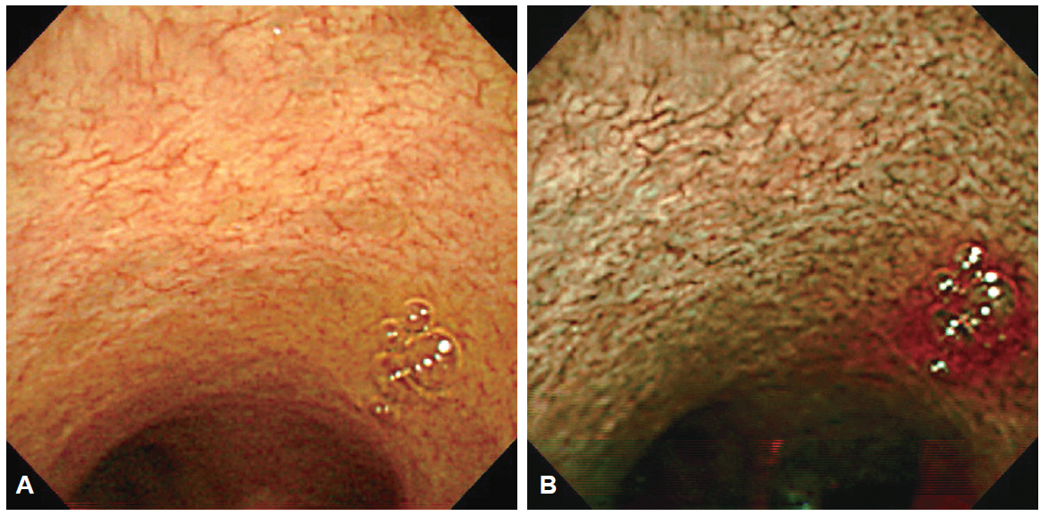
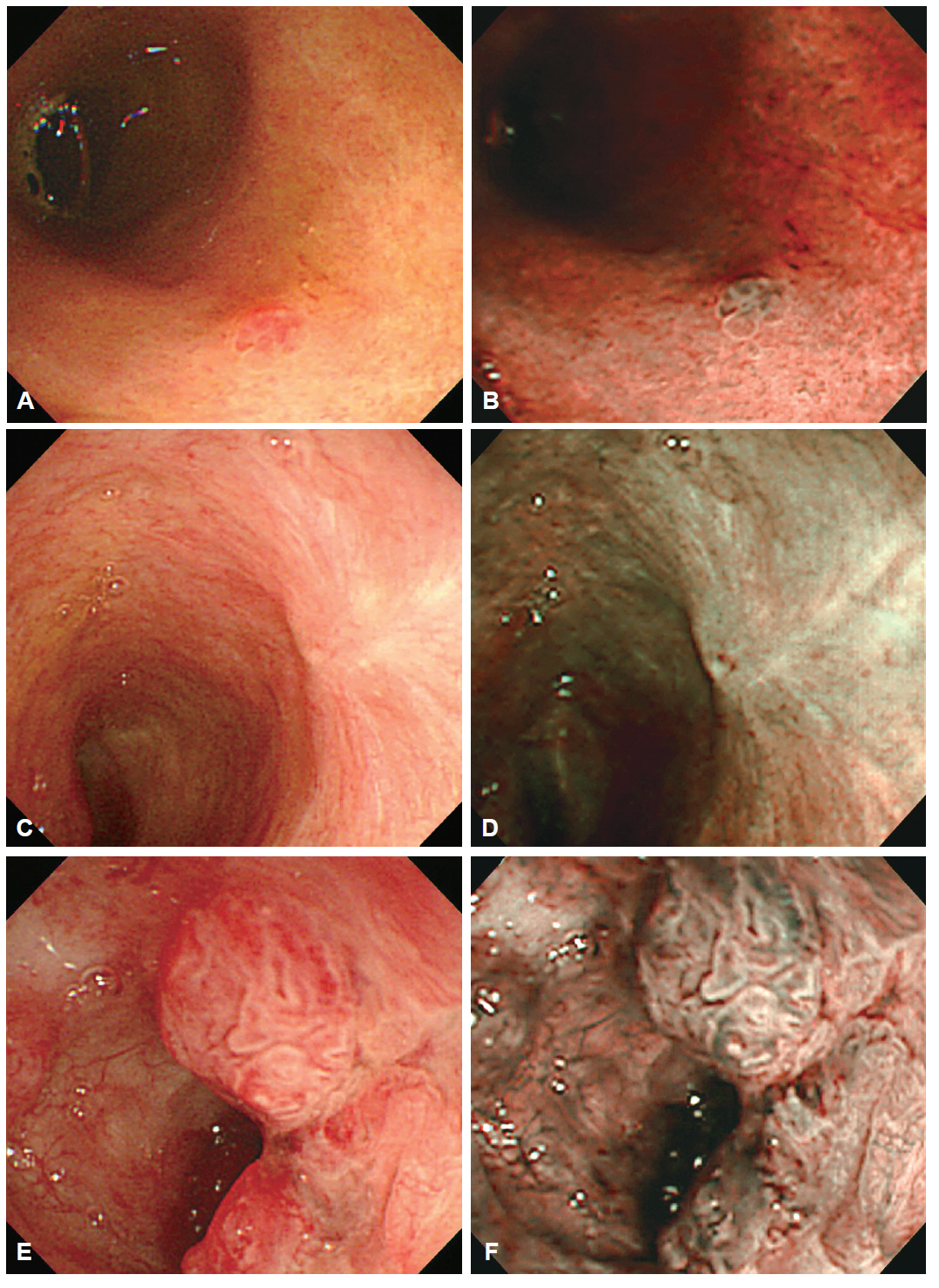
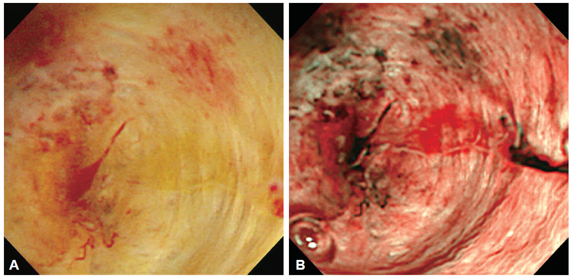
- Newly introduced direct peroral cholangioscopy and the development of video choledochoscopes have enabled more defined observation of bile duct mucosal lesions with clearer images. Narrow-band imaging (NBI) is a unique endoscopic imaging technology that provides enhanced endoscopic images of surface mucosal structures and its superficial microvessels. Advanced cholangioscopy and NBI are expected to be useful for precise evaluation and correct diagnosis of biliary tract diseases. However, the diagnostic value of advanced bile duct imaging with cholangioscopy requires further evaluation.
Figure
Reference
-
1. Larghi A, Waxman I. Endoscopic direct cholangioscopy by using an ultra-slim upper endoscope: a feasibility study. Gastrointest Endosc. 2006; 63:853–857.
Article2. Choi HJ, Moon JH, Ko BM, et al. Overtube-balloon-assisted direct peroral cholangioscopy by using an ultra-slim upper endoscope (with videos). Gastrointest Endosc. 2009; 69:935–940.
Article3. Moon JH, Ko BM, Choi HJ, et al. Intraductal balloon-guided direct peroral cholangioscopy with an ultraslim upper endoscope (with videos). Gastrointest Endosc. 2009; 70:297–302.
Article4. Moon JH, Terheggen G, Choi HJ, Neuhaus H. Peroral cholangioscopy: diagnostic and therapeutic applications. Gastroenterology. 2013; 144:276–282.
Article5. Itoi T, Sofuni A, Itokawa F, et al. Free-hand direct insertion ability into a simulated ex vivo model using a prototype multibending peroral direct cholangioscope (with videos). Gastrointest Endosc. 2012; 76:454–457.
Article6. Itoi T, Nageshwar Reddy D, Sofuni A, et al. Clinical evaluation of a prototype multi-bending peroral direct cholangioscope. Dig Endosc. 2014; 26:100–107.
Article7. Lee YN, Moon JH, Choi HJ, et al. A newly modified access balloon catheter for direct peroral cholangioscopy by using an ultraslim upper endoscope (with videos). Gastrointest Endosc. 2015 Aug 15 [Epub]. http://dx.doi.org/10.1016/j.gie.2015.08.021.8. Kim HJ, Kim MH, Lee SK, Yoo KS, Seo DW, Min YI. Tumor vessel: a valuable cholangioscopic clue of malignant biliary stricture. Gastrointest Endosc. 2000; 52:635–638.
Article9. Seo DW, Lee SK, Yoo KS, et al. Cholangioscopic findings in bile duct tumors. Gastrointest Endosc. 2000; 52:630–634.
Article10. Fukuda Y, Tsuyuguchi T, Sakai Y, Tsuchiya S, Saisyo H. Diagnostic utility of peroral cholangioscopy for various bile-duct lesions. Gastrointest Endosc. 2005; 62:374–382.
Article11. Kawakami H, Kuwatani M, Etoh K, et al. Endoscopic retrograde cholangiography versus peroral cholangioscopy to evaluate intraepithelial tumor spread in biliary cancer. Endoscopy. 2009; 41:959–964.
Article12. Osanai M, Itoi T, Igarashi Y, et al. Peroral video cholangioscopy to evaluate indeterminate bile duct lesions and preoperative mucosal cancerous extension: a prospective multicenter study. Endoscopy. 2013; 45:635–642.
Article13. Nishikawa T, Tsuyuguchi T, Sakai Y, et al. Preoperative assessment of longitudinal extension of cholangiocarcinoma with peroral video-cholangioscopy: a prospective study. Dig Endosc. 2014; 26:450–457.
Article14. Itoi T, Neuhaus H, Chen YK. Diagnostic value of image-enhanced video cholangiopancreatoscopy. Gastrointest Endosc Clin N Am. 2009; 19:557–566.
Article15. Ishida Y, Itoi T, Okabe Y. Can image-enhanced cholangioscopy distinguish benign from malignant lesions in the biliary duct? Best Pract Res Clin Gastroenterol. 2015; 29:611–625.
Article16. Itoi T, Sofuni A, Itokawa F, et al. Peroral cholangioscopic diagnosis of biliary-tract diseases by using narrow-band imaging (with videos). Gastrointest Endosc. 2007; 66:730–736.
Article17. Ishida Y, Okabe Y, Kaji R, et al. Evaluation of magnifying endoscopy using narrow band imaging using ex vivo bile duct (with video). Dig Endosc. 2013; 25:322–328.18. Azeem N, Gostout CJ, Knipschield M, Baron TH. Cholangioscopy with narrow-band imaging in patients with primary sclerosing cholangitis undergoing ERCP. Gastrointest Endosc. 2014; 79:773–779.
Article
- Full Text Links
- Actions
-
Cited
- CITED
-
- Close
- Share
- Similar articles
-
- Role of Image-Enhanced Endoscopy in Pancreatobiliary Diseases
- Early Bile Duct Cancer Detected by Direct Peroral Cholangioscopy with Narrow-Band Imaging after Bile Duct Stone Removal
- Advanced Imaging Technology Other than Narrow Band Imaging
- The Clinical Approach for Asymptomatic Bile Duct Dilatation
- Usefulness of Narrow-Band Imaging in Endoscopic Submucosal Dissection of the Stomach


