J Adv Prosthodont.
2017 Dec;9(6):482-485. 10.4047/jap.2017.9.6.482.
Biofilm formation on denture base resin including ZnO, CaO, and TiOâ‚‚ nanoparticles
- Affiliations
-
- 1Department of Prosthetic Dentistry, Regensburg University Medical Center, Regensburg, Germany. sebastian.hahnel@ukr.de
- KMID: 2398054
- DOI: http://doi.org/10.4047/jap.2017.9.6.482
Abstract
- PURPOSE
This laboratory study aimed to investigate the effect of doping an acrylic denture base resin material with nanoparticles of ZnO, CaO, and TiOâ‚‚ on biofilm formation.
MATERIALS AND METHODS
Standardized specimens of a commercially available cold-curing acrylic denture base resin material were doped with 0.1, 0.2, 0.4, or 0.8 wt% commercially available ZnO, CaO, and TiOâ‚‚ nanopowder. Energy dispersive X-ray spectroscopy (EDX) was used to identify the availability of the nanoparticles on the surface of the modified specimens. Surface roughness was determined by employing a profilometric approach; biofilm formation was simulated using a monospecies Candida albicans biofilm model and a multispecies biofilm model including C. albicans, Actinomyces naeslundii, and Streptococcus gordonii. Relative viable biomass was determined after 20 hours and 44 hours using a MTT-based approach.
RESULTS
No statistically significant disparities were identified among the various materials regarding surface roughness and relative viable biomass.
CONCLUSION
The results indicate that doping denture base resin materials with commercially available ZnO, CaO, or TiOâ‚‚ nanopowders do not inhibit biofilm formation on their surface. Further studies might address the impact of varying particle sizes as well as increasing the fraction of nanoparticles mixed into the acrylic resin matrix.
Keyword
MeSH Terms
Figure
-
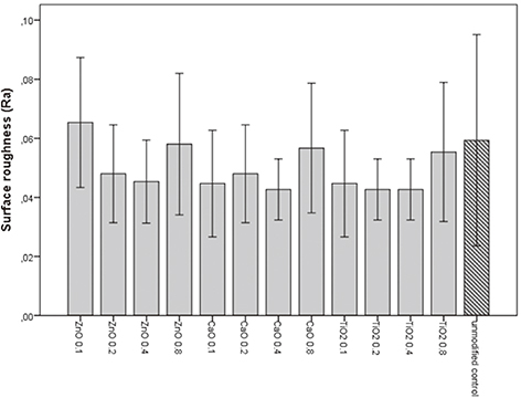
Fig. 1 Surface roughness (Ra) of the various modified denture base resins and the unmodified control. Means and standard deviations are indicated.

Fig. 2 Relative absorbance values indicating the relative amount of adherent viable cells in the monospecies biofilm model. Means and standard deviations are indicated.
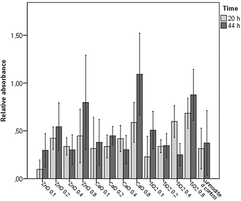
Fig. 3 Relative absorbance values indicating the relative amount of adherent viable cells in the multispecies biofilm model. Means and standard deviations are indicated.
Reference
-
1. Nitschke I, Stark H. Krankheits- und Versorgungspravalenzen bei jungeren Senioren (65- bis 74-Jahrige). In : Jordan AR, Micheelis W, editors. Funfte Deutsche Mundgesundheitsstudie. Cologne: Deutscher Zahnarzte Verlag DAV;2016. p. 416–451.2. Gendreau L, Loewy ZG. Epidemiology and etiology of denture stomatitis. J Prosthodont. 2011; 20:251–260.3. Budtz-Jorgensen E, Bertram U. Denture stomatitis I The etiology in relation to trauma and infection. Acta Odontol Scand. 1970; 28:71–92.4. Sawai J, Igarashi H, Hashimoto A, Kokugan T, Shimizu M. Evaluation of growth inhibitory effect of ceramics powder slurry on bacteria by conductance method. J Chem Eng Jpn. 1995; 28:288–293.5. Sawai J, Yoshikawa T. Quantitative evaluation of antifungal activity of metallic oxide powders (MgO, CaO and ZnO) by an indirect conductimetric assay. J Appl Microbiol. 2004; 96:803–809.6. Arai T, Ueda T, Sugiyama T, Sakurai K. Inhibiting microbial adhesion to denture base acrylic resin by titanium dioxide coating. J Oral Rehabil. 2009; 36:902–908.7. Azam A, Ahmed AS, Oves M, Khan MS, Habib SS, Memic A. Antimicrobial activity of metal oxide nanoparticles against Gram-positive and Gram-negative bacteria: a comparative study. Int J Nanomedicine. 2012; 7:6003–6009.8. Raghupathi KR, Koodali RT, Manna AC. Size-dependent bacterial growth inhibition and mechanism of antibacterial activity of zinc oxide nanoparticles. Langmuir. 2011; 27:4020–4028.9. Susewind S, Lang R, Hahnel S. Biofilm formation and Candida albicans morphology on the surface of denture base materials. Mycoses. 2015; 58:719–727.10. Cierech M, Kolenda A, Grudniak AM, Wojnarowicz J, Woźniak B, Gołaś M, Swoboda-Kopeć E, Łojkowski W, Mierzwińska-Nastalska E. Significance of polymethylmethacrylate (PMMA) modification by zinc oxide nanoparticles for fungal biofilm formation. Int J Pharm. 2016; 510:323–335.11. Dizaj SM, Lotfipour F, Barzegar-Jalali M, Zarrintan MH, Adibkia K. Antimicrobial activity of the metals and metal oxide nanoparticles. Mater Sci Eng C Mater Biol Appl. 2014; 44:278–284.
- Full Text Links
-
- Actions
-
Cited
- CITED
-
- Close
- Share
-
- Similar articles
-
- Accelerated ecotoxicity of photoreactive nanoparticles on Moina macrocopa
- A study of the antifungal properties and flexural strength of 3D printed denture base resin containing titanium dioxide nanoparticles
- Adhesion of biofilm, surface characteristics, and mechanical properties of antimicrobial denture base resin
- Preventive effects of shiitake mushroom extract on candida stomatitis
- In Vitro Studies on the Role of Zinc Oxide in the Development of Aspergillus Fumigatus Biofilm on Nasal Epithelial Cells
