Rare finding of Eustachian tube calcifications with cone-beam computed tomography
- Affiliations
-
- 1Department of Oral and Maxillofacial Medicine and Diagnostic Sciences, CWRU School of Dental Medicine, Cleveland, OH, USA. azs16@case.edu
- 2Senior Dental Student, CWRU School of Dental Medicine, Cleveland, OH, USA.
- 3Department of Comprehensive Care, CWRU School of Dental Medicine, Cleveland, OH, USA.
- KMID: 2397843
- DOI: http://doi.org/10.5624/isd.2017.47.4.275
Abstract
- Soft tissue calcification is a pathological condition in which calcium and phosphate salts are deposited in the soft tissue organic matrix. This study presents an unusual calcification noted in the cartilaginous portion of the Eustachian tube. A 67-year-old woman presented for dental treatment, specifically for implant placement, and cone-beam computed tomography (CBCT) was performed. The CBCT scan was reviewed by a board-certified oral and maxillofacial radiologist and revealed incidental findings of 2 distinct calcifications in the cartilaginous portion of the Eustachian tube. To the authors' knowledge, no previous study has reported the diagnosis of Eustachian tube calcification using CBCT. This report describes an uncommon variant of Eustachian tube calcification, which has a significant didactic value because such cases are seldom illustrated either in textbooks or in the literature. This case once again underscores the importance of having CBCT scans evaluated by a board-certified oral and maxillofacial radiologist.
MeSH Terms
Figure
-
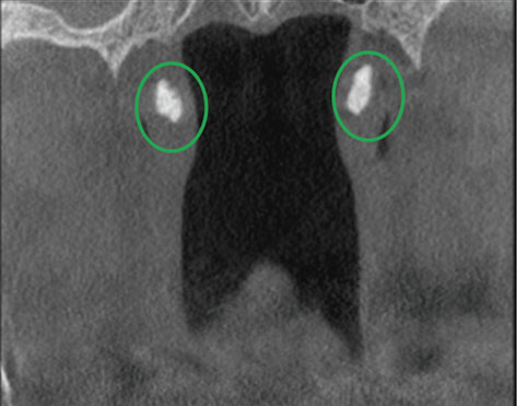
Fig. 1 Coronal cone-beam computed tomography image reveals bilateral calcifications (circles) of the cartilaginous Eustachian tube.
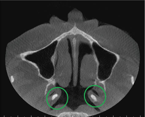
Fig. 2 Axial cone-beam computed tomography image depicts the bilateral calcifications (circles) of the cartilaginous Eustachian tube.
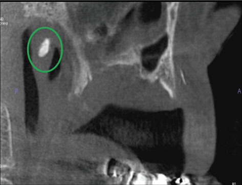
Fig. 3 Sagittal cone-beam computed tomography image depicts the bilateral calcifications (circles) of the cartilaginous Eustachian tube.
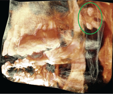
Fig. 4 Volume rendering demonstrates the Eustachian tube calcifications (circle).
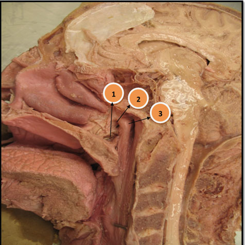
Fig. 5 Midsagittal section of a cadaver showing the nasopharynx region and antoamical landmarks surrounding the Eustachian tube. 1. Tensor veli palatini muscle insertion on the soft palate. 2. Opening of the Eustachian tube. 3. Torus tubarius.
Cited by 1 articles
-
Eustachian Tube Function Test
Jeon Mi Lee, Hyun Jin Lee
Korean J Otorhinolaryngol-Head Neck Surg. 2022;65(4):193-201. doi: 10.3342/kjorl-hns.2022.00220.
Reference
-
1. Syed AZ, Mupparapu M. Fossa navicularis magna detection on cone-beam computed tomography. Imaging Sci Dent. 2016; 46:47–51.
Article2. Syed AZ, Sin C, Rios R, Mupparapu M. Incidental occurrence of an unusually large mastoid foramen on cone-beam computed tomography and review of the literature. Imaging Sci Dent. 2016; 46:39–45.
Article3. Syed AZ, Zahedpasha S, Rathore SA, Mupparapu M. Evaluation of canalis basilaris medianus using cone-beam computed tomography. Imaging Sci Dent. 2016; 46:141–144.
Article4. Sardesai V, Gharpuray M. Calcinosis cutis. Indian J Dermatol Venereol Leprol. 2003; 69:45–46.5. Hussmann J, Russell RC, Kucan JO, Khardori R, Steinau HU. Soft-tissue calcifications: differential diagnosis and therapeutic approaches. Ann Plast Surg. 1995; 34:138–147.6. Lanka P, Lanka LR, Ethirajan N, Krishnaswamy B, Manohar U. Idiopathic calcinosis cutis. Indian J Dermatol. 2009; 54:388–389.
Article7. Takasaki K, Sando I, Balaban CD, Haginomori S, Ishijima K, Kitagawa M. Histopathological changes of the eustachian tube cartilage and the tensor veli palatini muscle with aging. Laryngoscope. 1999; 109:1679–1683.
Article8. Buch K, Nadgir RN, Qureshi MM, Ozonoff A, Sakai O. Clinical significance of incidentally detected torus tubarius calcification. J Comput Assist Tomogr. 2017; 41:828–832.
Article9. Alper CM, Swarts JD, Singla A, Banks J, Doyle WJ. Relationship between the electromyographic activity of the paratubal muscles and eustachian tube opening assessed by sonotubometry and videoendoscopy. Arch Otolaryngol Head Neck Surg. 2012; 138:741–746.
Article10. Takasaki K, Takahashi H, Miyamoto I, Yoshida H, Yamamoto-Fukuda T, Enatsu K, et al. Measurement of angle and length of the eustachian tube on computed tomography using the multiplanar reconstruction technique. Laryngoscope. 2007; 117:1251–1254.
Article11. Leuwer R. Anatomy of the Eustachian tube. Otolaryngol Clin North Am. 2016; 49:1097–1106.
Article12. Bluestone CD, Bluestone MB. Eustachian tube: structure, function, role in otitis media. Hamilton: BC Decker;2005.13. Morshedi MM, Mafee MF. Calcification of the cartilaginous Eustachian tube. Neuroradiology. 2012; 54:525–527.
Article14. Rosenfeld RM, Bluestone CD. Evidence-based otitis media. 2nd Ed. Hamilton: BC Decker;2003.15. Smith ME, Scoffings DJ, Tysome JR. Imaging of the Eustachian tube and its function: a systematic review. Neuroradiology. 2016; 58:543–556.
Article16. Moore KL, Dalley AF, Agur AM. Clinically oriented anatomy. 7th Ed. Philadelphia: Lippincott Williams & Wilkins Health;2014.17. Turk LM, Hogg DA. Age changes in the human laryngeal cartilages. Clin Anat. 1993; 6:154–162.
Article18. White SC, Pharoah MJ. Oral radiology: principles and interpretation. 7th Ed. St. Louis: Elsevier Mosby;2014.19. Takasaki K, Thompson SW, Sando I. Ossification of eustachian tube cartilage and Ostmann's fatty tissue in chronic renal failure. Otolaryngol Head Neck Surg. 2000; 122:567–571.
Article
- Full Text Links
-
- Actions
-
Cited
- CITED
-
- Close
- Share
-
- Similar articles
-
- Eustachian tube calcification as an unusual finding on a panoramic radiograph
- Management of root canal perforation by using cone-beam computed tomography
- A rare case of dilated invaginated odontome with talon cusp in a permanent maxillary central incisor diagnosed by cone beam computed tomography
- Multiple intraosseous cervical pneumatocysts: A case report of a rare incidental finding on cone-beam computed tomography
- Cone beam computed tomography findings of ectopic mandibular third molar in the mandibular condyle: report of a case
