Multispectral X-ray imaging to distinguish among dental materials
- Affiliations
-
- 1Department of Prosthodontics and Orofacial Function, School of Dentistry, Philipps-University Marburg/Lahn, Germany. gente@med.uni-marburg.de
- KMID: 2397839
- DOI: http://doi.org/10.5624/isd.2017.47.4.247
Abstract
- PURPOSE
Dual-energy X-ray imaging is widely used today in various areas of medicine and in other applications. However, no similar technique exists for dental applications. In this study, we propose a dual-energy technique for dental diagnoses based on voltage-switching.
MATERIALS AND METHODS
The method presented in this study allowed different groups of materials to be classified based on atomic number, thereby enabling two-dimensional images to be colorized. Computer simulations showed the feasibility of this approach. Using a number of different samples with typical biologic and synthetic dental materials, the technique was applied to radiographs acquired with a commercially available dental X-ray unit.
RESULTS
This technique provided a novel visual representation of the intraoral environment in three colors, and is of diagnostic value when compared to state-of-the-art grayscale images, since the oral cavity often contains multiple permanent foreign materials.
CONCLUSION
This work developed a technique for two-dimensional dual-energy imaging in the context of dental applications and showed its feasibility with a commercial dental X-ray unit in simulation and experimental studies.
MeSH Terms
Figure
-
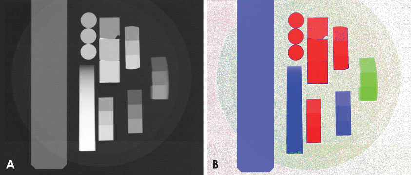
Fig. 1 Comparison of classical grayscale and new colored radiographs. The proposed method assigned distinct colors to different materials based on their atomic number. (A) Classical X-ray image, (B) New dual-energy analyis.
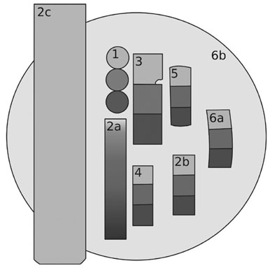
Fig. 2 Material composition of the first sample holder. Samples were glued onto a polymethyl methacrylate (PMMA) disc that was held by an aluminum rod from the X-ray unit (2c). To investigate the influence of material thickness on subsequent analysis, different sample dimensions were included.
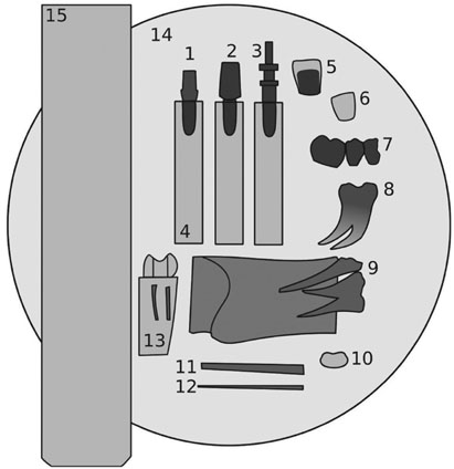
Fig. 3 Schematic drawing of the second sample holder: 1. Implant; 2. Implant with abutment (PEEK); 3. Implant; 4. PMMA; 5. Ceramic crown; 6. Veneer; 7. Gold bridge; 8. Tooth; 9. Teeth of a pig with bone; 10. Sealer; 11 and 12. Gutta-percha points; 13. Tooth in , polymethyl methacrylate (PMMA); 14. PMMA; 15. Aluminum. PMMA, polymethyl methacrylate.

Fig. 4 Schematic drawing of the third sample holder. 1. Guttaflow; 2. Apexit; 3. Exp. Ormocer LC; 4. GrandioSo; 5. RealSeal; 6. Gutta-percha points; 7. Zirconium; 8. Lower jaw of a pig; 9. AH Plus; 10. Aluminum; 11. polymethyl methacrylate (PMMA).
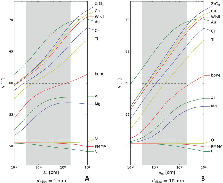
Fig. 5 Results of the computer simulation. The quantity h was evaluated for different materials and varying sample thicknesses dm. The thicknesses of interest for dental applications are shaded in gray. Dotted horizontal lines separate groups of materials with similar properties. The thickness of the PMMA disc ddisc was assumed to be 2 mm (A) or 15 mm (B).
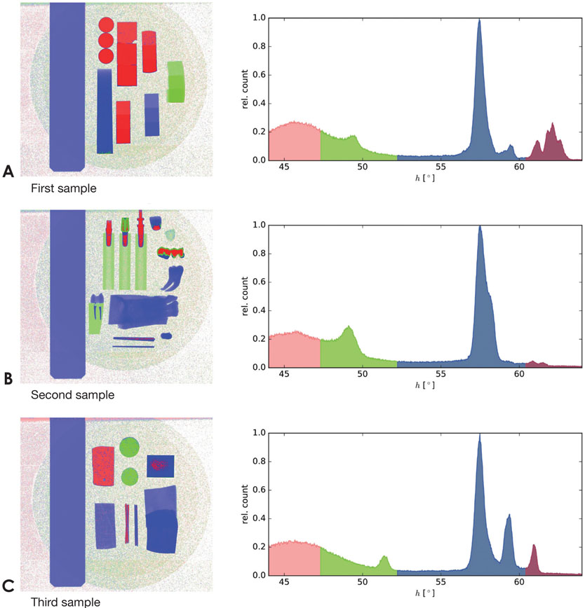
Fig. 6 Experimental results. The panels A, B, and C show the joined colored X-ray images (left) with corresponding histograms of the value of h (right). Depending on the value of h, four discrete material classes could be identified. Panel A displays the results from the first sample holder, panel B the results from the second sample holder and panel C the results from the third sample holder.
Reference
-
1. Alvarez RE, Macovski A. Energy-selective reconstructions in X-ray computerized tomography. Phys Med Biol. 1976; 21:733–744.2. Macovski A, Alvarez RE, Chan JL, Stonestrom JP, Zatz LM. Energy dependent reconstruction in X-ray computerized tomography. Comput Biol Med. 1976; 6:325–336.
Article3. Genant HK, Boyd D. Quantitative bone mineral analysis using dual energy computed tomography. Invest Radiol. 1977; 12:545–551.
Article4. Krane KS, Halliday D. Introductory nuclear physics. New York: Wiley;1988.5. Rutherford RA, Pullan BR, Isherwood I. Measurement of effective atomic number and electron density using an EMI scanner. Neuroradiology. 1976; 11:15–21.
Article6. Friedmann H. Einführung in die Kernphysik. Berlin: Wiley-VCH;2014.7. Simons D, Kachelriess M, Schlemmer HP. Recent developments of dual-energy CT in oncology. Eur Radiol. 2014; 24:930–939.
Article8. Petersilka M, Bruder H, Krauss B, Stierstorfer K, Flohr TG. Technical principles of dual source CT. Eur J Radiol. 2008; 68:362–368.
Article9. Flohr TG, McCollough CH, Bruder H, Petersilka M, Gruber K, Süss C, et al. First performance evaluation of a dual-source CT (DSCT) system. Eur Radiol. 2006; 16:256–268.
Article10. Johnson TR, Krauss B, Sedlmair M, Grasruck M, Bruder H, Morhard D, et al. Material differentiation by dual energy CT: initial experience. Eur Radiol. 2007; 17:1510–1517.
Article11. Taibi A, Fabbri S, Baldelli P, di Maggio C, Gennaro G, Marziani M, et al. Dual-energy imaging in full-field digital mammography: a phantom study. Phys Med Biol. 2003; 48:1945–1956.
Article12. Jochelson MS, Dershaw DD, Sung JS, Heerdt AS, Thornton C, Moskowitz CS, et al. Bilateral contrast-enhanced dual-energy digital mammography: feasibility and comparison with conventional digital mammography and MR imaging in women with known breast carcinoma. Radiology. 2013; 266:743–751.
Article13. Meyer BC, Werncke T, Hopfenmüller W, Raatschen HJ, Wolf KJ, Albrecht T. Dual energy CT of peripheral arteries: effect of automatic bone and plaque removal on image quality and grading of stenoses. Eur J Radiol. 2008; 68:414–422.
Article14. Apfaltrer P, Sudarski S, Schneider D, Nance JW Jr, Haubenreisser H, Fink C, et al. Value of monoenergetic low-kV dual energy CT datasets for improved image quality of CT pulmonary angiography. Eur J Radiol. 2014; 83:322–328.
Article15. Marin D, Nelson RC, Barnhart H, Schindera ST, Ho LM, Jaffe TA, et al. Detection of pancreatic tumors, image quality, and radiation dose during the pancreatic parenchymal phase: effect of a low-tube-voltage, high-tube-current CT technique - preliminary results. Radiology. 2010; 256:450–459.16. Adams JE, Chen SZ, Adams PH, Isherwood I. Measurement of trabecular bone mineral by dual energy computed tomography. J Comput Assist Tomogr. 1982; 6:601–607.
Article17. Deslattes RD, Kessler EG Jr, Indelicato P, de Billy L, Lindroth E, Anton J. X-ray transition energies: new approach to a comprehensive evaluation. Rev Mod Phys. 2003; 75:35–99.
Article18. Brunetti A, del Rio MS, Golosio B, Simionovici A, Somogyi A. A library for X-ray-matter interaction cross sections for X-ray fluorescence applications. Spectrochim Acta Part B At Spectrosc. 2004; 59:1725–1731.
Article19. Schoonjans T, Brunetti A, Golosio B, del Rio MS, Solé VA, Ferrero C, et al. The xraylib library for X-ray-matter interactions. Recent developments. Spectrochim Acta Part B At Spectrosc. 2011; 66:776–784.
Article20. Pasler FA. Zahnärztliche Radiologie. Stuttgart: Georg Thieme Verlag;2008.
- Full Text Links
-
- Actions
-
Cited
- CITED
-
- Close
- Share
-
- Similar articles
-
- Unwanted effects due to interactions between dental materials and magnetic resonance imaging: a review of the literature
- Leakage and scattered radiation from hand-held dental x-ray unit
- Effect of the amount of battery charge on tube voltage in different hand-held dental x-ray systems
- Message from the President of the Korean Society of Nuclear Medicine
- Absorbed and effective dose for periapical radiography using portable and wall type dental X-ray machines
