Intraoperative Transcranial Sonography for Detection of Contralateral Hematoma Volume Change in Patients with Traumatic Brain Injury
- Affiliations
-
- 1Department of Neurosurgery, Medical Research Institute, Pusan National University Hospital, Pusan National University School of Medicine, Busan, Korea. bc1743kim@gmail.com
- KMID: 2394548
- DOI: http://doi.org/10.13004/kjnt.2017.13.2.137
Abstract
- The authors present two clinical cases, in which intraoperative transcranial sonography (TCS) was used to detect a change in contralateral hematoma volume. A 51-year-old female and a 5-year-old male underwent osteoplastic craniotomy for epidural hematoma removal. Scant contralateral hematoma was evident by preoperative computed tomography in both patients. Intraoperative TCS was used to detect changes in contralateral hematomas. After observing a volume change in one case, a second operation was performed immediately. Based in this experience, the authors recommend intraoperative TCS for the detection of contralateral hematoma volume changes.
MeSH Terms
Figure
-
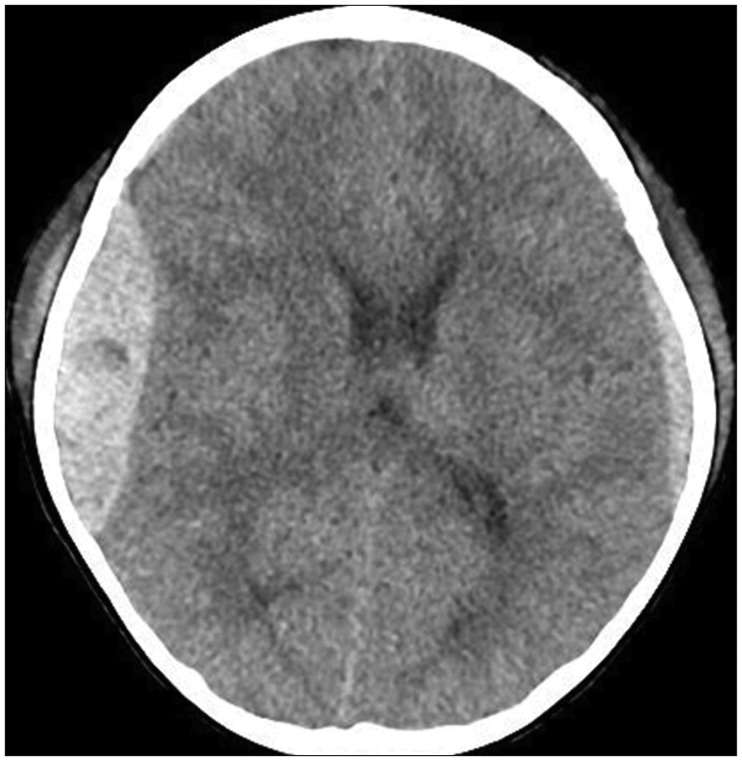
FIGURE 1 A 5-year-old man admitted after a fall from a height of 2 m. The initial computed tomography scan showed an epidural hematoma on right temporoparietal area and subdural hematoma on the contralateral side.
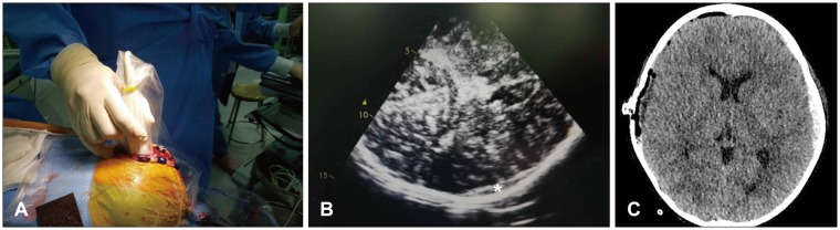
FIGURE 2 (A) Intraoperative transcranial sonography (TCS) was performed. (B) After evacuation of the right temporoparietal hematoma, intraoperative TCS showed no changed subdural hematoma (asterisk) on the contralateral side. (C) Postoperative computed tomography confirmed an unchanged subdural hematoma status on the contralateral side.
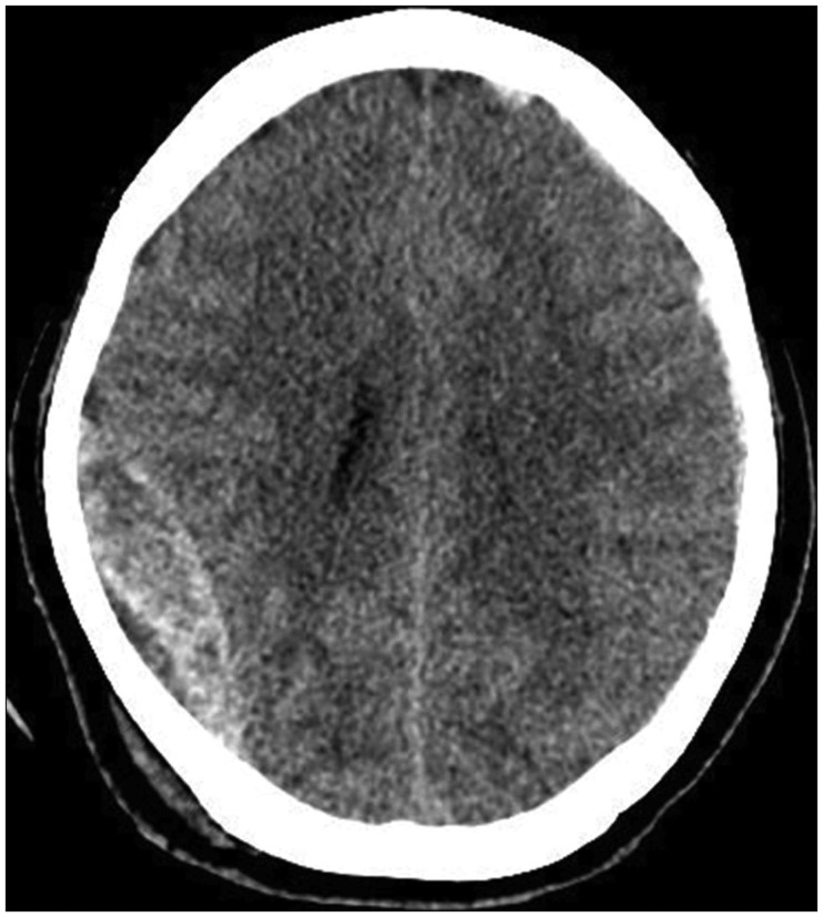
FIGURE 3 A 51-year-old female admitted after a pedestrian accident. Initial computed tomography scan showed an epidural hematoma on the right temporoparietal area and subdural hematoma on the left frontotemporal area.
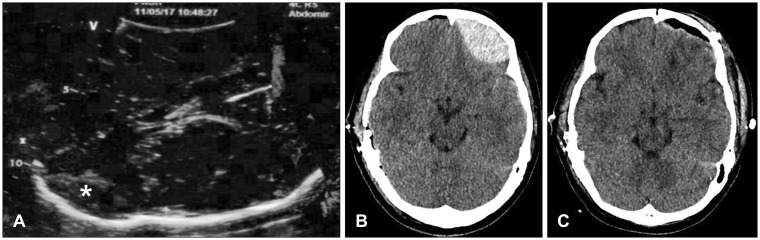
FIGURE 4 (A) After evacuation of right temporoparietal hematoma, intraoperative transcranial sonography showed a newly developed epidural hematoma (EDH) (asterisk) on the left frontal area. (B) Postoperative computed tomography confirmed newly developed EDH on the left frontal area. (C) EDH on the left frontal area was evacuated by secondary operation.
Reference
-
1. Aaslid R, Markwalder TM, Nornes H. Noninvasive transcranial Doppler ultrasound recording of flow velocity in basal cerebral arteries. J Neurosurg. 1982; 57:769–774. PMID: 7143059.
Article2. Becker G, Berg D, Rausch WD, Lange HK, Riederer P, Reiners K. Increased tissue copper and manganese content in the lentiform nucleus in primary adult-onset dystonia. Ann Neurol. 1999; 46:260–263. PMID: 10443894.
Article3. Becker G, Seufert J, Bogdahn U, Reichmann H, Reiners K. Degeneration of substantia nigra in chronic Parkinson's disease visualized by transcranial color-coded real-time sonography. Neurology. 1995; 45:182–184. PMID: 7824114.
Article4. Borovich B, Braun J, Guilburd JN, Zaaroor M, Michich M, Levy L, et al. Delayed onset of traumatic extradural hematoma. J Neurosurg. 1985; 63:30–34. PMID: 4009271.
Article5. Boviatsis EJ, Korfias S, Kouyialis AT, Sakas DE. Epidural haematoma after evacuation of contralateral subdural haematoma. Ir J Med Sci. 2004; 173:217–218. PMID: 16323618.
Article6. Bucci MN, Phillips TW, McGillicuddy JE. Delayed epidural hemorrhage in hypotensive multiple trauma patients. Neurosurgery. 1986; 19:65–68. PMID: 3748339.
Article7. Caricato A, Pitoni S, Montini L, Bocci MG, Annetta P, Antonelli M. Echography in brain imaging in intensive care unit: State of the art. World J Radiol. 2014; 6:636–642. PMID: 25276307.
Article8. Duffner F, de Zwart P, Bitzer M, Will B. Subacute and chronic epidural hematoma after craniocerebral trauma. Aktuelle Traumatol. 1993; 23:345–349. PMID: 8147252.9. Kern R, Kablau M, Sallustio F, Fatar M, Stroick M, Hennerici MG, et al. Improved detection of intracerebral hemorrhage with transcranial ultrasound perfusion imaging. Cerebrovasc Dis. 2008; 26:277–283. PMID: 18648201.
Article10. Niesen WD, Burkhardt D, Hoeltje J, Rosenkranz M, Weiller C, Sliwka U. Transcranial grey-scale sonography of subdural haematoma in adults. Ultraschall Med. 2006; 27:251–255. PMID: 16596509.11. Oh MJ, Jeong JH, Shin DS, Hwang SC, Im SB, Kim BT, et al. Postoperative contralateral hematoma in patient with acute traumatic brain injury. Korean J Neurotrauma. 2017; 13:24–28. PMID: 28512614.
Article12. Ostrup R, Bejar R, Marshall L. Real-time ultrasonography: a useful tool in the evaluation of the craniectomized, brain-injured patient. Neurosurgery. 1983; 12:225–227. PMID: 6835507.
Article13. Pozzati E, Frank F, Frank G, Gaist G. Subacute and chronic extradural hematomas: a study of 30 cases. J Trauma. 1980; 20:795–799. PMID: 7411669.14. Saberi H, Meybodi AT, Meybodi KT, Habibi Z, Mirsadeghi SM. Delayed post-operative contralateral epidural hematoma in a patient with right-sided acute subdural hematoma: a case report. Cases J. 2009; 2:6282. PMID: 19918570.
Article15. Santa M, Sulla I, Fagul'a J, Santová I. Two-dimensional ultrasonographic examination through postoperative defects in skull. Zentralbl Neurochir. 1990; 51:194–196. PMID: 2099055.
- Full Text Links
-
- Actions
-
Cited
- CITED
-
- Close
- Share
-
- Similar articles
-
- Usefulness of intraoperative transcranial sonography in patients with traumatic brain injuries: a comparison with postoperative computed tomography
- Intraoperative Development of Contralateral Subdural Hematoma during Evacuation of Acute Subdural Hematoma: Case Report
- Postoperative Contralateral Hematoma in Patient with Acute Traumatic Brain Injury
- Contralateral Epidural Hematoma Developing after the Craniotomy for Traumatic Intracranial Hematoma.
- Delirium After Traumatic Brain Injury: Prediction by Location and Size of Brain Lesion
