Obstet Gynecol Sci.
2017 Sep;60(5):481-484. 10.5468/ogs.2017.60.5.481.
Metastatic uterine cancer looking as cervical fibroid in recurrent breast cancer woman: a case report
- Affiliations
-
- 1Department of Obstetrics and Gynecology, Inje University Haeundae Paik Hospital, Inje University College of Medicine, Busan, Korea. jyimdog@paik.ac.kr
- 2Department of General Surgery, Inje University Haeundae Paik Hospital, Inje University College of Medicine, Busan, Korea.
- KMID: 2393850
- DOI: http://doi.org/10.5468/ogs.2017.60.5.481
Abstract
- Metastasis to the female genital tract from extragenital primary cancer is uncommon. In this case, a 46-year-old woman was diagnosed with invasive lobular carcinoma of the left breast in 2011. She had left breast conserving surgery, chemotherapy, radiation, and hormonal therapy (gosereline and tamoxifen). However, she developed menorrhagia after interruption of hormonal therapy and incidentally, the ultrasonogram of her pelvis showed a solid, large mass in the cervix. It looked like leiomyoma. Because of massive vaginal bleeding requiring multiple blood transfusions, she underwent total hysterectomy with bilateral salpingo-oophorectomy. Unexpectedly, however, histopathological examination revealed metastatic carcinoma, consistent with breast origin.The metastatic tumor involved the uterine corpus with spreading to the endocervix, left ovary, and multiple lymphovascular invasion was present. We described the rarity and risk of metastatic uterine cancer in patient with history of malignant tumor treatment.
Keyword
MeSH Terms
Figure
-
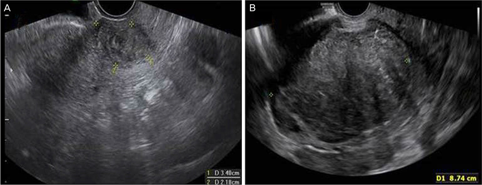
Fig. 1 (A) Vaginal ultrasonography showing a tumor of the cervix (uteri, 3.4 cm in diameter). (B) The cervix appears as a large, cervical leiomyoma (8.7 cm in 2017).

Fig. 2 (A) The cut surface of the bisected uterus in the direction from anterior to posterior shows a well demarcated, solid, mass-like lesion; the lesion is yellow, it contains necrotic focus (arrow). (B) Tumor cells lack cohesion and appear individually dispersed through a collagenous stroma, and they occasionally have a signet-ring cell appearance with intracytoplasmic mucin.
Reference
-
1. Kamby C, Vejborg I, Kristensen B, Olsen LO, Mouridsen HT. Metastatic pattern in recurrent breast cancer. Special reference to intrathoracic recurrences. Cancer. 1988; 62:2226–2233.2. Pomerance W, MacKles A. Adenocarcinoma of the cervix. Am J Obstet Gynecol. 1962; 84:367–374.3. Green AE, Biscotti C, Michener C, Belinson J. Isolated cervical metastasis of breast cancer: a case report and review of the literature. Gynecol Oncol. 2004; 95:267–269.4. Lokadasan R, Ratheesan K, Sukumaran R, Nair SP. Metastatic lobular carcinoma of breast mimics primary cervix carcinoma: two case reports and a review of the literature. Ecancermedicalscience. 2015; 9:571.5. Abell MR, Gosling JR. Gland cell carcinoma (adenocarcinoma) of the uterine cervix. Am J Obstet Gynecol. 1962; 83:729–755.6. Piura B, Yanai-Inbar I, Rabinovich A, Zalmanov S, Goldstein J. Abnormal uterine bleeding as a presenting sign of metastases to the uterine corpus, cervix and vagina in a breast cancer patient on tamoxifen therapy. Eur J Obstet Gynecol Reprod Biol. 1999; 83:57–61.7. Pestalozzi BC, Zahrieh D, Mallon E, Gusterson BA, Price KN, Gelber RD, et al. Distinct clinical and prognostic features of infiltrating lobular carcinoma of the breast: combined results of 15 International Breast Cancer Study Group clinical trials. J Clin Oncol. 2008; 26:3006–3014.8. Sastre-Garau X, Jouve M, Asselain B, Vincent-Salomon A, Beuzeboc P, Dorval T, et al. Infiltrating lobular carcinoma of the breast. Clinicopathologic analysis of 975 cases with reference to data on conservative therapy and metastatic patterns. Cancer. 1996; 77:113–120.
- Full Text Links
-
- Actions
-
Cited
- CITED
-
- Close
- Share
-
- Similar articles
-
- A Case of Breast Cancer Metastatic Solely to the Uterine Cervix
- Metastatic Breast Cancer from Cervical Cancer
- Unusual Peritoneal Metastasis of Late Recurrent Uterine Cervical Cancer: A Case Report and Literature Review
- A Case of Colonic Metastasis of Recurrent Cervical Cancer
- A Case of Recurred Uterine Cervical Cancer Presented as Only Huge Mediastinal Mass
