Shear bond strength of veneering porcelain to zirconia and metal cores
- Affiliations
-
- 1Department of Prosthodontics, Graduate School, Seoul National University, Seoul, Korea. proshan@snu.ac.kr
Abstract
- STATEMENT OF PROBLEM: Zirconia-based restorations have the common technical complication of delamination, or porcelain chipping, from the zirconia core. Thus the shear bond strength between the zirconia core and the veneering porcelain requires investigation in order to facilitate the material's clinical use. PURPOSE: The purpose of this study was to evaluate the bonding strength of the porcelain veneer to the zirconia core and to other various metal alloys (high noble metal alloy and base metal alloy). MATERIAL AND METHODS: 15 rectangular (4x4x9mm) specimens each of zirconia (Cercon), base metal alloy (Tillite), high noble metal alloy (Degudent H) were fabricated for the shear bond strength test. The veneering porcelain recommended by the manufacturer for each type of material was fired to the core in thickness of 3mm. After firing, the specimens were embedded in the PTFE mold, placed on a mounting jig, and subjected to shear force in a universal testing machine. Load was applied at a crosshead speed of 0.5mm/min until fracture. The average shear strength (MPa) was analyzed with the one-way ANOVA and the Tukey's test (alpha= .05). The fractured specimens were examined using SEM and EDX to determine the failure pattern. RESULTS: The mean shear strength (+/- SD) in MPa was 25.43 (+/- 3.12) in the zirconia group, 35.87 (+/- 4.23) in the base metal group, 38.00 (+/- 5.23) in the high noble metal group. The ANOVA showed a significant difference among groups, and the Tukey's test presented a significant difference between the zirconia group and the metal group. Microscopic examination showed that the failure primarily occurred near the interface with the residual veneering porcelain remaining on the core. CONCLUSION: There was a significant difference between the metal ceramic and zirconia ceramic group in shear bond strength. There was no significant difference between the base metal alloy and the high noble metal alloy.
MeSH Terms
Figure
-

Fig. 1 Final dimensions of specimens.
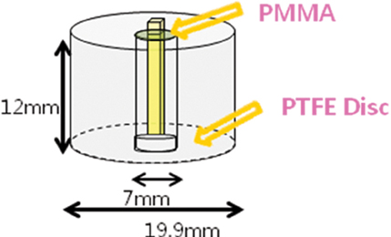
Fig. 2 Schematic diagram of specimen embedded in PTFE molds.

Fig. 3 Schematic diagram of SBS test.

Fig. 4 SEM image of zirconia-veneer group (Group I). (A) The arrow indicates the direction of load. The loaded side demonstrates cohesive failure within the veneering porcelain (original magnification × 30), (B) Note many pores within veneering 'porcelain (arrow), where fracture originated. The fractured Cercon Ceram kiss veneer demonstrates multiple cracks extending in a vertical direction (Hackle patterns) (original magnification × 250), (C) High magnification SEM image exhibited a very thin layer of porcelain covering zirconia grains (original magnification × 1000).

Fig. 5 SEM image of base metal alloy-veneer group (Group II). (A) The arrow indicates the direction of load. The loaded side demonstrates cohesive failure within the veneering porcelain (original magnification × 30), (B) Interface of the veneering porcelain and the metal core (original magnification × 250), (C) High magnification SEM image exhibited an opaque layer and an oxide layer (original magnification × 1000).

Fig. 6 SEM image of high noble metal alloy-veneer group (Group III) (A) The arrow indicates the direction of load (original magnification × 30), (B) Predominance of cohesive failure (original magnification × 250), (C) High magnification SEM image exhibited an opaque layer and oxide layer (original magnification × 1000).

Fig. 7 EDX results of Group I, II, III. (A) EDX results of Group I showed the presence of a thin porcelain layer over the zirconia, (B) EDX results of the fractured base metal alloy surface (Group II) demonstrated an exposed metal surface with some ceramic remainder, (C) EDX results of the fractured high noble metal alloy surface (Group III) presented no exposed metal surface.
Reference
-
1. Tan K, Pjetursson BE, Lang NP, Chan ES. A systemic review of the survival and complication rates of fixed partial dentures (FPDs) after an observation period of at least 5 years. Clin Oral Implants Res. 2004. 15:654–666.2. Raigrodski AJ. Contemporary materials and technologies for all-ceramic fixed partial dentures: a review of the literature. J Prosthet Dent. 2004. 92:557–562.3. Guazzato M, Albakry M, Ringer SP, Swain MV. Strength, fracture toughness and microstructure of a selection of all-ceramic materials. Part II. Zirconia based dental ceramics. Dent Mater. 2004. 20:449–456.4. Tinschert J, Natt G, Mohrbotter N, Spiekermann H, Schulze KA. Lifetime of alumina- and zircoina ceramics used for crown and bridge restorations. J Biomed Mater Res B Appl Biomater. 2007. 80:317–321.5. Sundh A, Sjogren G. A comparison of fracture strength of yttrium-oxide-partially-stabilized zirconia ceramic crowns with varying core thickness, shapes and veneer ceramics. J Oral Rehabil. 2004. 31:682–688.6. Sailer I, Pjetursson BE, Zwahlen M, Hammerle CHF. A Systematic review of the survival and complication rates of all-ceramic and metal-ceramic reconstructions after an observation period of at least 3 years. Part II: Fixed partial prostheses. Clin Oral Implants Res. 2007. 18:86–96.7. Sailer I, Feher A, Filser F. Prospective clinical study of zirconia posterior fixed partial dentures: 3-year follow-up. Quintessence Int. 2006. 37:685–693.8. Raigrodski AJ, Chiche GJ, Potiket N. The efficacy of posterior three-unit zirconium-oxide-based ceramic fixed partial denture prostheses: a prospective clinical pilot study. J Prosthet Dent. 2006. 96:237–244.9. Guazzato M, Proos K, Sara G, Swain MV. Strength, reliability, and mode of fracture of bilayered porcelain/core ceramics. Int J Prosthodont. 2004. 17:142–149.10. Anusavice KJ. Phillips' science of dental materials. 2003. 11th Ed. Philadelphia: W.B. Saunders;621–654.11. Ozcan M, Niedermeier W. Clinical study on the reasons and location of the failures of metal-ceramic restorations and survival of repairs. Int J Prosthodont. 2002. 15:299–302.12. Craig RG, Powers JM. Restorative dental materials. 2002. 11th ed. Mosby.13. ISO 9693 Metal-ceramic bond characterization (Schwickerath crack initiation test). 1999. Geneva, Switzerland: International Organization for Standardization.14. Isgro G, Pallav P, van der Zel JM, Feilzer AJ. The influence of the veneering porcelain and different surface treatments on the biaxial flexural strength of a heat-pressed ceramic. J Prosthet Dent. 2003. 90:465–473.15. Hara AT, Pimenta LA, Rodrigues AL Jr. Influence of crosshead speed on resin-dentin shear bond strength. Dent Mater. 2001. 17:165–169.16. Albakry M, Guazzato M, Swain MV. Fracture toughness and hardness evaluation of three pressable all-ceramic dental materials. J Dent. 2003. 31:181–188.17. Al-Dohan HM, Yaman P, Dennison JB, Razzoog ME, Lang BR. Shear strength of core-veneer interface in bi-layered ceramics. J Prosthet Dent. 2004. 91:349–355.18. Dündar M, Ozcan M, Cömlekoglu E, Güngör MA, Artunç C. Bond strengths of veneering ceramics to reinforced ceramic core materials. Int J Prosthodont. 2005. 18:71–72.19. Dündar M, Ozcan M, Gökçe B, Cömlekoglu E, Leite F, Valandro LF. Comparison of two bond strength testing methodologies for bilayered all-ceramics. Dent Mater. 2007. 23:630–636.20. Guess PC, Kulis A, Witkowski S, Wolkewitz M, Zhang Y, Strub JR. Shear bond strengths between different zirconia cores and veneering ceramics and their susceptibility to thermocycling. Dent Mater. 2008. 24:1556–1567.21. White SN, Miklus VG, McLaren EA, Lang LA, Caputo AA. Flexural strength of a layered zirconia and porcelain dental all-ceramic system. J Prosthet Dent. 2005. 94:125–131.22. Aboushelib MN, de Jager N, Kleverlaan CJ, Feilzer AJ. Microtensile bond strength of different components of core veneered all-ceramic restorations. Dent Mater. 2005. 21:984–991.23. Aboushelib MN, Kleverlaan CJ, Feiler AJ. Microtensile bond strength of different components of core veneered all-ceramic restorations. Part II: zirconia veneering ceramics. Dent Mater. 2006. 22:857–863.24. de Melo RM, Travassos AC, Neisser MP. Shear bond strengths of a ceramic system to alternative metal alloys. J Prosthet Dent. 2005. 93:64–69.25. Mecholsky J. Fractography: Determining the sites of fracture initiation. Dent Mater. 1995. 11:113–116.26. Kelly JR, Giordano R, Pober R, Cima MJ. Fracture surface analysis of dental ceramics: clinically failed restorations. Int J Prosthodont. 1990. 3:430–440.27. Quinn JB, Sundar V, Lloyd IK. Influence of microstructure and chemistry on the fracture toughness of dental ceramics. Dent Mater. 2003. 19:603–611.28. Aboushelib MN, Kleverlaan CJ, Feilzer AJ. Microtenisile bond strength of different componentsof core veneered all-ceramic restorations. Part 3: double veneer tenchnique. J Prosthodont. 2008. 17:9–13.29. Sobrinho LC, Cattell MJ, Glover RH, Knowles JC. Investigation of the dry and wet fatique properties of three all-ceramic crown systems. Int J Prosthodont. 1998. 11:255–262.
- Full Text Links
-
- Actions
-
Cited
- CITED
-
- Close
- Share
-
- Similar articles
-
- Shear bond strength of veneer ceramic and colored zirconia by using aqueous metal chloride solutions
- A study on the shear bond strengths of veneering ceramics to the colored zirconia core
- Effect of surface treatmet on the shear bond strength of a zirconia core to veneering ceramic
- Evaluation of shear bond strengths of gingiva-colored composite resin to porcelain, metal and zirconia substrates
- Shear bond strength of veneering ceramic to coping materials with different pre-surface treatments
