J Bone Metab.
2014 Feb;21(1):2-7.
Quality Control of DXA System and Precision Test of Radio-technologists
- Affiliations
-
- 1Department of Nuclear Medicine, Asan Medical Center, Ulsan University School of Medicine, Seoul, Korea.
- 2Department of Nuclear Medicine (Radiology), Dongnam Institute of Radiological and Medical Science, Busan, Korea. soyangmd@naver.com
Abstract
- The image quality management of bone mineral density (BMD) is the responsibility and duty of radio-technologists who carry out examinations. However, inaccurate conclusions due to the lack of understanding and ignorance regarding the methodology of image quality management can be a fatal error to patients. The accuracy and precision of BMD measurement must be maintained at the highest level so that actual biological changes can be detected with even slight changes in BMD. Accuracy and precision should be continuously preserved for image quality of machines. Those factors will contribute to ensure the reliability of BMD examination. The enforcement of proper quality control of radiologists performing BMD inspections which brings about the durability extensions of equipment and accurate results of calculations will help the assurance of reliable inspections.
Figure
-

Fig. 1 General phantoms and scan images. (A) The European Spine Phantom (ESP; QRM GmbH Dorfstrasse 4. 91096, Möhrendorf, Germany). (B) ESP scan image. (C) Bio-imaging bone fide spine phantom (BFP; Bio-Imaging Technologies Inc., Newtown, PA, USA). (D) BFP scan image.
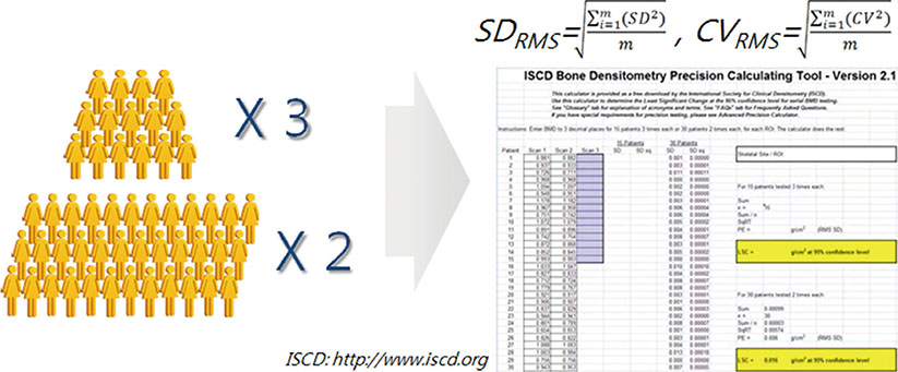
Fig. 2 Precision test method.

Fig. 3 Calculation of least significant change (LSC) at 95% confidence level (in case of device error: 1%, operator error: 1%).
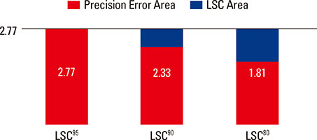
Fig. 4 Variation of precision error according to confidence level.
Reference
-
1. Lenchik L, Leib ES, Hamdy RC, et al. Executive summary International Society for Clinical Densitometry position development conference Denver, Colorado July 20-22, 2001. J Clin Densitom. 2002; 5:S1–S3.
Article2. Kim DY. Clinical application of bone mineral density measurement. Korean J Nucl Med. 2004; 38:275–281.3. Faulkner KG, McClung MR. Quality control of DXA instruments in multicenter trials. Osteoporos Int. 1995; 5:218–227.
Article4. Fuerst TP. Quality control of dual X-ray absorptiometry systems. In : Gould RG, Boone JM, editors. RSNA categorical course in physics. Oak Brook, IL: Radiological Society of North America;1996. p. 67–76.5. Blunt BA, Jones D, Svensen RK, et al. Good clinical practice and audits for dual X-ray absorptiometry and X-ray imaging laboratories and quality assurance centers involved in clinical drug trials, private practice, and research. J Clin Densitom. 1998; 1:323–337.
Article6. Orwoll ES, Oviatt SK. The Nafarelin/Bone Study Group. Longitudinal precision of dual-energy x-ray absorptiometry in a multicenter study. J Bone Miner Res. 1991; 6:191–197.
Article7. Montgomery DC. Introduction to statistical quality control. 3rd ed. New York, NY: John Wiley & Sons;1996.8. Orwoll ES, Oviatt SK, Biddle JA. Precision of dual-energy x-ray absorptiometry: development of quality control rules and their application in longitudinal studies. J Bone Miner Res. 1993; 8:693–699.
Article9. Lu Y, Mathur AK, Blunt BA, et al. Dual X-ray absorptiometry quality control: comparison of visual examination and process-control charts. J Bone Miner Res. 1996; 11:626–637.
Article10. Faulkner KG, Gluer CC, Estilo M, et al. Cross-calibration of DXA equipment: upgrading from a Hologic QDR 1000/W to a QDR 2000. Calcif Tissue Int. 1993; 52:79–84.
Article11. British Standards Institution. Guide to data analysis and quality control using cusum techniques: BS 5703. London, UK: British Standards Institution;1980.12. Pearson D, Cawte SA. Long-term quality control of DXA: a comparison of Shewhart rules and Cusum charts. Osteoporos Int. 1997; 7:338–343.
Article13. Garland SW, Lees B, Stevenson JC. DXA longitudinal quality control: a comparison of inbuilt quality assurance, visual inspection, multi-rule Shewhart charts and Cusumanalysis. Osteoporos Int. 1997; 7:231–237.
Article14. Bonnick SL, Johnston CC Jr, Kleerekoper M, et al. Importance of precision in bone density measurements. J Clin Densitom. 2001; 4:105–110.
Article15. The Writing Group for the ISCD Position Development Conference. Technical standardization for dual-energy x-ray absorptiometry. J Clin Densitom. 2004; 7:27–36.16. The International Society for Clinical Densitometry. The precision calculating tool. 2013. cited by 2013 January 1. Available from: http://www.iscd.org/.17. Gluer CC, Blake G, Lu Y, et al. Accurate assessment of precision errors: how to measure the reproducibility of bone densitometry techniques. Osteoporos Int. 1995; 5:262–270.
Article18. Miller PD, Bonmick SL. Clinical application of bone densitometry. In : Falus MJ, editor. Primer in the metabolic bone diseases and disorders of mineral metabolism. 4th ed. Philadelphia, PA: Lippincott Williams and Wilkins;1999. p. 158.19. Lenchik L, Kiebzak GM, Blunt BA. What is the role of serial bone mineral density measurements in patient management? J Clin Densitom. 2002; 5:S29–S38.
Article
- Full Text Links
-
- Actions
-
Cited
- CITED
-
- Close
- Share
-
- Similar articles
-
- Clinical Application of Bone Mineral Density Measurement
- Measurement and Interpretation of Dual-Energy X-ray Absorptiometry Bone Density Measurements
- The Level of Medical Technologists' Perception of and Compliance with Hospital Infection Control Guidelines
- The Evaluation of the OPUS(R) Immunoassay System for the Serum CK-MB Test
- Lyophilized Quality Control Material for Clinical Chemistry and Coagulation
