Infect Chemother.
2013 Mar;45(1):99-104.
Endobronchial Mycobacterium avium Infection in an Immunocompetent Patient
- Affiliations
-
- 1Department of Internal Medicine, Korea Cancer Center Hospital, Korea Institute of Radiological and Medical Sciences, Seoul, Korea. slowly7@kcch.re.kr
- 2Department of Pathology, Korea Cancer Center Hospital, Korea Institute of Radiological and Medical Sciences, Seoul, Korea.
Abstract
- Although Mycobacterium avium complex (MAC) is the most common pathogen in nontuberculous mycobacterial (NTM) pulmonary diseases, endobronchial lesions caused by MAC infections are very rare even in an immunocompromised host. Herein, we describe the case of a 59-year-old, HIV-negative and non-immunocompromised woman who developed multifocal pulmonary infiltrations with endobronchial lesion caused by M. avium. Bronchoscopic examination revealed white- and yellow-colored irregular mucosal lesions in the bronchus of the left lingular division. M. avium was identified using sputum culture and bronchial washing fluid culture. Following the recommendations of the American Thoracic Society and Infectious Diseases Society of America (ATS/IDSA), the patient was begun on treatment with antimycobacterial drugs. After treatment, pneumonic infiltration decreased.
MeSH Terms
Figure
-
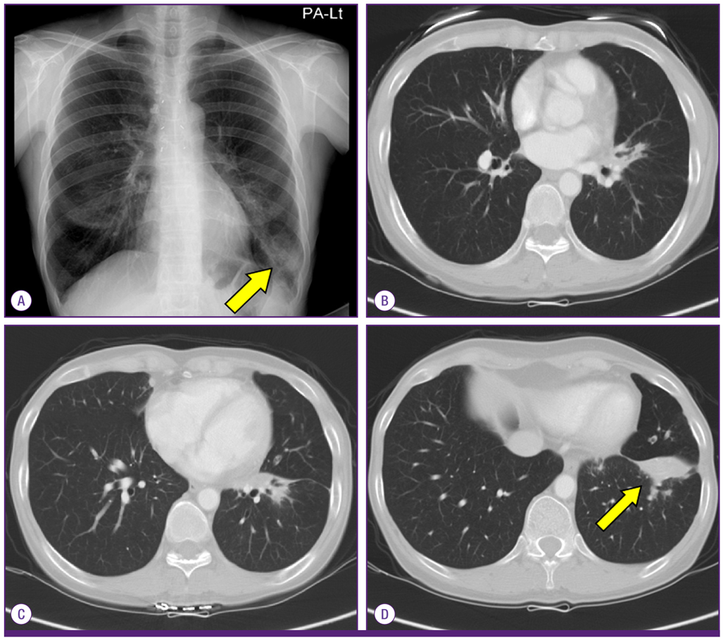
Figure 1 Chest radiography (A) and chest CT (B-D) at the initial visit showing multiple pulmonary infiltrates in the left lingular division and left lower lobe.

Figure 2 Chest radiography (A) and chest CT (B-D) after 6 months showing aggravation of the multiple pulmonary infiltrates in the left lingular division and left lower lobe.
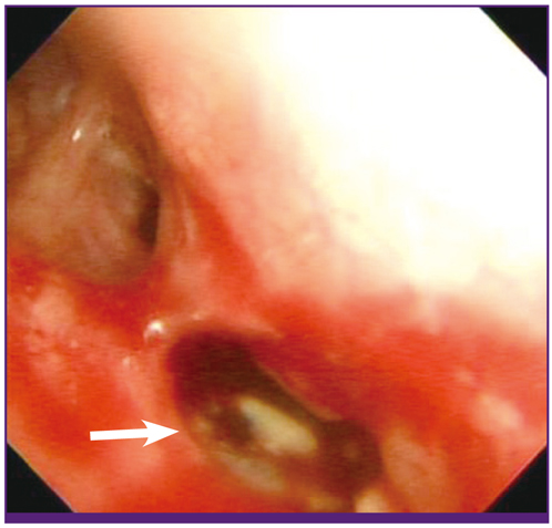
Figure 3 Bronchoscopic view from the bronchus of the left lingular division shows a white- and yellow-colored irregular mucosal lesion.
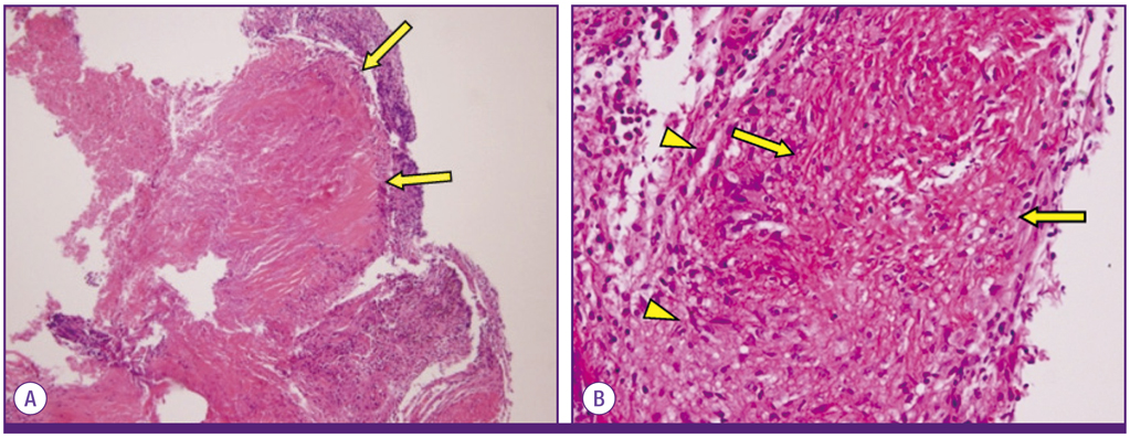
Figure 4 Microscopic finding of chronic granulomatous inflammation with caseous necrosis. (A) H&E staining at 100× magnification; (B) H&E staining at 400× magnification; arrow, caseous necrosis; arrowhead, epithelioid histiocyte.
Reference
-
1. Griffith DE, Aksamit T, Brown-Elliott BA, Catanzaro A, Daley C, Gordin F, Holland SM, Horsburgh R, Huitt G, Iademarco MF, Iseman M, Olivier K, Ruoss S, von Reyn CF, Wallace RJ Jr, Winthrop K. ATS Mycobacterial Diseases Subcommittee. American Thoracic Society. Infectious Disease Society of America. An official ATS/IDSA statement: diagnosis, treatment, and prevention of nontuberculous mycobacterial diseases. Am J Respir Crit Care Med. 2007. 175:367–416.
Article2. Koh WJ, Kwon OJ, Lee KS. Diagnosis and treatment of non-tuberculous mycobacterial pulmonary diseases: a Korean perspective. J Korean Med Sci. 2005. 20:913–925.
Article3. Shih JY, Wang HC, Chiang IP, Yang PC, Luh KT. Endobronchial lesions in a non-AIDS patient with disseminated Mycobacterium avium-intracellulare infection. Eur Respir J. 1997. 10:497–499.
Article4. Asano T, Itoh G, Itoh M. Disseminated Mycobacterium intracellulare infection in an HIV-negative, nonimmunosuppressed patient with multiple endobronchial polyps. Respiration. 2002. 69:175–177.
Article5. Fukuoka K, Nakano Y, Nakajima A, Hontsu S, Kimura H. Endobronchial lesions involved in Mycobacterium avium infection. Respir Med. 2003. 97:1261–1264.
Article6. Manali ED, Tomford WJ, Liao DW, Farver C, Mehta AC. Mycobacterium kansasii endobronchial ulcer in a nonimmunocompromised patient. Respiration. 2005. 72:305–308.
Article7. del Rio Camacho G, Soriano Guillén L, Flandes Aldeyturriaga J, Hernández García B, Bernácer Borja M. Endobronchial atypical mycobacteria in an immunocompetent child. Pediatr Pulmonol. 2010. 45:511–513.
Article8. Litman DA, Shah UK, Pawel BR. Isolated endobronchial atypical mycobacterium in a child: a case report and review of the literature. Int J Pediatr Otorhinolaryngol. 2000. 55:65–68.
Article9. Lee JH, Son KS, Park JH, Kim JC, Lee HW, Kim CH. Mycobacterium avium infection presenting as endobronchial lesions in an immunocompetent patient. Tuberc Respir Dis. 2006. 60:571–575.
Article10. Lee YJ, Kim DH, Yoon KH, Kim MY, Jung SW, Lee BK, Kim YJ. A case of pulmonary and endobronchial Mycobacterium avium infection presenting as an acute pneumonia in an immunocompetent patient. Tuberc Respir Dis. 2010. 69:279–283.
Article11. Park YS, Lee CH, Lee SM, Yang SC, Yoo CG, Kim YW, Han SK, Shim YS, Yim JJ. Rapid increase of non-tuberculous mycobacterial lung diseases at a tertiary referral hospital in South Korea. Int J Tuberc Lung Dis. 2010. 14:1069–1071.12. Chen CY, Chen HY, Chou CH, Huang CT, Lai CC, Hsueh PR. Pulmonary infection caused by nontuberculous mycobacteria in a medical center in Taiwan, 2005-2008. Diagn Microbiol Infect Dis. 2012. 72:47–51.
Article13. Chung HS, Lee JH. Bronchoscopic assessment of the evolution of endobronchial tuberculosis. Chest. 2000. 117:385–392.
Article14. Kashyap S, Mohapatra PR, Saini V. Endobronchial tuberculosis. Indian J Chest Dis Allied Sci. 2003. 45:247–256.15. Smart J. Endobronchial tuberculosis. Br J Dis Chest. 1951. 45:61–68.
Article
- Full Text Links
-
- Actions
-
Cited
- CITED
-
- Close
- Share
-
- Similar articles
-
- A Case of Pulmonary and Endobronchial Mycobacterium avium Infection Presenting as an Acute Pneumonia in an Immunocompetent Patient
- Mycobacterium avium Infection Presenting as Endobronchial Lesions in an Immunocompetent Patient
- Mycobacterium Avium Complex Infection Presenting as an Endobronchial Mass in a Patient with Acquired Immune Deficiency Syndrome
- Vertebral Osteomyelitis due to Mycobacterium intracellulare in an Immunocompetent Elderly Patient After Vertebroplasty
- Isolated Endobronchial Mycobacterium avium Disease Associated with Lobar Atelectasis in an Immunocompetent Young Adult: A Case Report and Literature Review
