Imaging Sci Dent.
2017 Mar;47(1):1-9. 10.5624/isd.2017.47.1.1.
Pharyngeal airway dimensions in skeletal class II: A cephalometric growth study
- Affiliations
-
- 1Clinic of Orthodontics, Ministry of Health, Tepebası Oral and Dental Health Hospital, Ankara, Turkey. ozgeusluakcam@gmail.com
- KMID: 2391400
- DOI: http://doi.org/10.5624/isd.2017.47.1.1
Abstract
- PURPOSE
This retrospective study aimed to evaluate the nasopharyngeal and oropharyngeal dimensions of individuals with skeletal class II, division 1 and division 2 patterns during the pre-peak, peak, and post-peak growth periods for comparison with a skeletal class I control group.
MATERIALS AND METHODS
Totally 124 lateral cephalograms (47 for skeletal class I; 45 for skeletal class II, division 1; and 32 for skeletal class II, division 2) in pre-peak, peak, and post-peak growth periods were selected from the department archives. Thirteen landmarks, 4 angular and 4 linear measurements, and 4 proportional calculations were obtained. The ANOVA and Duncan test were applied to compare the differences among the study groups during the growth periods.
RESULTS
Statistically significant differences were found between the skeletal class II, division 2 group and other groups for the gonion-gnathion/sella-nasion angle. The sella-nasion-B-point angle was different among the groups, while the A-point-nasion-B-point angle was significantly different for all 3 groups. The nasopharyngeal airway space showed a statistically significant difference among the groups throughout the growth periods. The interaction among the growth periods and study groups was statistically significant regarding the upper oropharyngeal airway space measurement. The lower oropharyngeal airway space measurement showed a statistically significant difference among the groups, with the smallest dimension observed in the skeletal class II, division 2 group.
CONCLUSION
The naso-oropharyngeal airway dimensions showed a statistically significant difference among the class II, division 1; class II, division 2; and class I groups during different growth periods.
Keyword
Figure
-

Fig. 1 Hand-wrist radiographs of the patients in the pre-peak (A), peak (B), and post-peak (C) growth period groups.

Fig. 2 Cephalometric radiographs of a skeletal class II patient in pre-peak (A), peak (B), and post-peak (C) growth period groups.
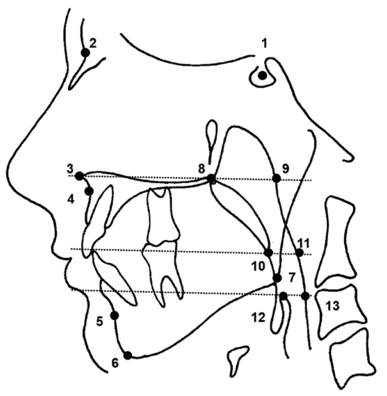
Fig. 3 Cephalometric landmarks are shown on a traced image of a sample class I case. For definitions, see Table 2.
Reference
-
1. McNamara JA. Influence of respiratory pattern on craniofacial growth. Angle Orthod. 1981; 51:269–300. PMID: 6947703.
Article2. Han S, Choi YJ, Chung CJ, Kim JY, Kim KH. Long-term pharyngeal airway changes after bionator treatment in adolescents with skeletal Class II malocclusions. Korean J Orthod. 2014; 44:13–19. PMID: 24511511.
Article3. Soni J, Shyagali TR, Bhayya DP, Shah R. Evaluation of pharyngeal space in different combinations of Class II skeletal malocclusion. Acta Inform Med. 2015; 23:285–289. PMID: 26635436.
Article4. Zinsly SR, Moraes LC, Moura P, Ursi W. Assessment of pharyngeal airway space using cone-beam computed tomograpy. Dental Press J Orthod. 2010; 15:150–158.5. Martin SE, Mathur R, Marshall I, Douglas NJ. The effect of age, sex, obesity and posture on upper airway size. Eur Respir J. 1997; 10:2087–2090. PMID: 9311508.
Article6. Tourné LP. Growth of the pharynx and its physiologic implications. Am J Orthod Dentofacial Orthop. 1991; 99:129–139. PMID: 1990822.
Article7. Johnston CD, Richardson A. Cephalometric changes in adult pharyngeal morphology. Eur J Orthod. 1999; 21:357–362. PMID: 10502898.
Article8. Martin O, Muelas L, Vinas MJ. Nasopharyngeal cephalometric study of ideal occlusions. Am J Orthod Dentofacial Orthop. 2006; 130:436.e1–436.e9. PMID: 17045141.
Article9. Späth-Schwalbe E, Hundenborn C, Kern W, Fehm HL, Born J. Nocturnal wakefulness inhibits growth hormone (GH)-releasing hormone-induced GH secretion. J Clin Endocrinol Metab. 1995; 80:214–219. PMID: 7829614.
Article10. Born J, Muth S, Fehm HL. The significance of sleep onset and slow wave sleep for nocturnal release of growth hormone (GH) and cortisol. Psychoneuroendocrinology. 1988; 13:233–243. PMID: 3406323.
Article11. Bollhalder J, Hanggi MP, Schatzle M, Markic G. Dentofacial and upper airway characteristics of mild and severe class II division 1 subjects. Eur J Orthod. 2013; 35:447–453. PMID: 22427406.
Article12. Battagel JM, Johal A, Kotecha B. A cephalometric comparison of subjects with snoring and obstructive sleep apnoea. Eur J Orthod. 2000; 22:353–365. PMID: 11029825.
Article13. Lowe AA, Fleetham JA, Adachi S, Ryan CF. Cephalometric and computed tomographic predictors of obstructive sleep apnoea severity. Am J Orthod Dentofacial Orthop. 1995; 107:589–595. PMID: 7771363.14. Yavuz B, Kocadereli İ. Sınıf II malokluzyonlarda uygulanan tedavi yaklaşımlarının üst hava yolu üzerine etkileri. EÜ Dişhek Fak Derg. 2013; 34:66–72.15. Coben SE. The biology of Class II treatment. Am J Orthod. 1971; 59:470–487. PMID: 5279861.
Article16. Souki BQ, Pimenta GB, Souki MQ, Franco LP, Becker HM, Pinto JA. Prevalence of malocclusion among mouth breathing children: do expectations meet reality? Int J Pediatr Otorhinolaryngol. 2009; 73:767–773. PMID: 19282036.
Article17. Agren K, Nordlander B, Linder-Aronsson S, Zettergren-Wijk L, Svanborg E. Children with nocturnal upper airway obstruction: postoperative orthodontic and respiratory improvement. Acta Otolaryngol. 1998; 118:581–587. PMID: 9726687.18. Kim YJ, Hong JS, Hwang YI, Park YH. Three-dimensional analysis of pharyngeal airway in preadolescent children with different anteroposterior skeletal patterns. Am J Orthod Dentofacial Orthop. 2010; 137:306.e1–306.e11. PMID: 20197163.
Article19. Güngör OE, Celikoğlu M, Kale B, Güngör AY, Sarı Z. The reliability of the Greulich and Pyle atlas when applied to a Southern Turkish population. Eur J Dent. 2015; 9:251–254. PMID: 26038659.
Article20. Jakhi SA, Karjodkar FR. Use of cephalometry in diagnosing resonance disorders. Am J Orthod Dentofacial Orthop. 1990; 98:323–332. PMID: 2220693.
Article21. Wu JT, Huang GF, Huang CS, Noordhoff MS. Nasopharyngoscopic evaluation and cephalometric analysis of velopharynx in normal and cleft palate patients. Ann Plast Surg. 1996; 36:117–123. PMID: 8919372.
Article22. Aboudara C, Nielsen I, Huang JC, Maki K, Miller AJ, Hatcher D. Comparison of airway space with conventional lateral headfilms and 3-dimensional reconstruction from cone-beam computed tomography. Am J Orthod Dentofacial Orthop. 2009; 135:468–479. PMID: 19361733.
Article23. El H, Palomo JM. Airway volume for different dentofacial skeletal patterns. Am J Orthod Dentofacial Orthop. 2011; 139:e511–e521. PMID: 21640863.
Article24. Hellsing E. Changes in pharyngeal airway in relation to extension of the head. Eur J Orthod. 1989; 11:359–365. PMID: 2591483.25. Pracharktam N, Hans MG, Strohl KP, Redline S. Upright and supine cephalometric evaluation of obstructive sleep apnoea syndrome an snoring subjects. Angle Orthod. 1994; 64:63–73. PMID: 8172396.26. Handelman CS, Osborne G. Growth of the nasopharynx and adenoid development from one to eighteeen years. Angle Orthod. 1976; 46:243–259. PMID: 1066976.27. Jeans WD, Fernando DC, Maw AR, Leighton BC. A longitudinal study of the growth of the nasopharynx and its contents in normal children. Br J Radiol. 1981; 54:117–121. PMID: 7459548.
Article28. Subtelny JD. Malocclusions, orthodontic corrections and orofacial muscle adaptation. Angle Orthod. 1970; 40:170–201. PMID: 5269951.29. Ceylan I, Oktay H. A study on the pharyngeal size in different skeletal patterns. Am J Orthod Dentofacial Orthop. 1995; 108:69–75. PMID: 7598107.
Article30. Zhong Z, Tang Z, Gao X, Zeng XL. A comparison study of upper airway among different skeletal craniofacial patterns in nonsnoring Chinese children. Angle Orthod. 2010; 80:267–274. PMID: 19905851.
Article31. Sosa FA, Graber TM, Muller TP. Postpharyngeal lymphoid tissue in Angle Class I and Class II malocclusions. Am J Orthod. 1982; 81:299–309. PMID: 6960717.
Article32. Wenzel A, Williams S, Ritzau M. Relationships of changes in craniofacial morphology, head posture, and nasopharyngeal airway size following mandibular osteotomy. Am J Orthod Dentofacial Orthop. 1989; 96:138–143. PMID: 2756949.
Article33. de Freitas MR, Alcazar NM, Janson G, de Freitas KM, Henriques JF. Upper and lower pharyngeal airways in subjects with Class I and Class II malocclusions and different growth patterns. Am J Orthod Dentofacial Orthop. 2006; 130:742–745. PMID: 17169736.
Article34. Alves PV, Zhao L, O'Gara M, Patel PK, Bolognese A. Three-dimensional cephalometric study of upper airway space in skeletal class II and III healthy patients. J Craniofac Surg. 2008; 19:1497–1507. PMID: 19098539.
Article35. Memon S, Fida M, Shaikh A. Comparison of different craniofacial patterns with pharyngeal widths. J Coll Physicians Surg Pak. 2012; 22:302–306. PMID: 22538035.36. Kerr WJ. The nasopharynx, face height and overbite. Angle Orthod. 1985; 55:31–36. PMID: 3856405.37. Keçik D. The effect of mandibular position on upper airway dimensions. Turk J Orthod. 2009; 22:93–101.38. Grauer D, Cevidanes LS, Styner MA, Ackerman JL, Proffit WR. Pharyngeal airway volume and shape from cone-beam computed tomography: relationship to facial morphology. Am J Orthod Dentofacial Orthop. 2009; 136:805–814. PMID: 19962603.
Article39. Ozbek MM, Memikoglu TU, Gögen H, Lowe AA, Baspinar E. Oropharyngeal airway dimensions and functional-orthopedic treatment in skeletal Class II cases. Angle Orthod. 1998; 68:327–336. PMID: 9709833.
- Full Text Links
-
- Actions
-
Cited
- CITED
-
- Close
- Share
-
- Similar articles
-
- Pharyngeal Airway Dimensions in Skeletal Class II Young Adolescents : Cephalometric Study
- Long-term pharyngeal airway changes after bionator treatment in adolescents with skeletal Class II malocclusions
- Does surgically assisted maxillary protraction with skeletal anchorage and Class III elastics affect the pharyngeal airway? A retrospective, long-term study
- Comparison of the effects on the pharyngeal airway space of maxillary protraction appliances according to the methods of anchorage
- Effect of extraction treatment on upper airway dimensions in patients with bimaxillary skeletal protrusion relative to their vertical skeletal pattern
