J Korean Soc Surg Hand.
2017 Sep;22(3):189-195. 10.12790/jkssh.2017.22.3.189.
Surgical Treatment of Metacarpal and Phalangeal Fracture with Rotational Malalignment
- Affiliations
-
- 1Department of Orthopaedic Surgery, CHA Bundang Medical Center, CHA University School of Medicine, Seongnam, Korea. hsoohong@hanmail.net
- 2Department of Orthopaedic Surgery, Seoul Paik Hospital, Inje University, Seoul, Korea.
- KMID: 2391216
- DOI: http://doi.org/10.12790/jkssh.2017.22.3.189
Abstract
- PURPOSE
Hand fractures can be treated conservatively in many cases, but rotation malalignment is one of the important indications for surgical treatment because of dysfunction. We performed open reduction and internal fixation in these malalignment fractures and report clinical and radiological results.
METHODS
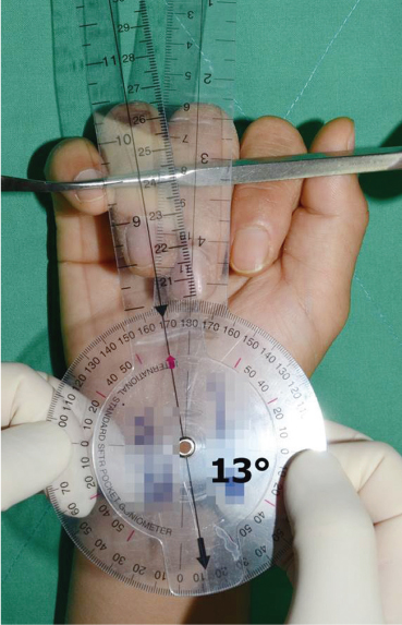
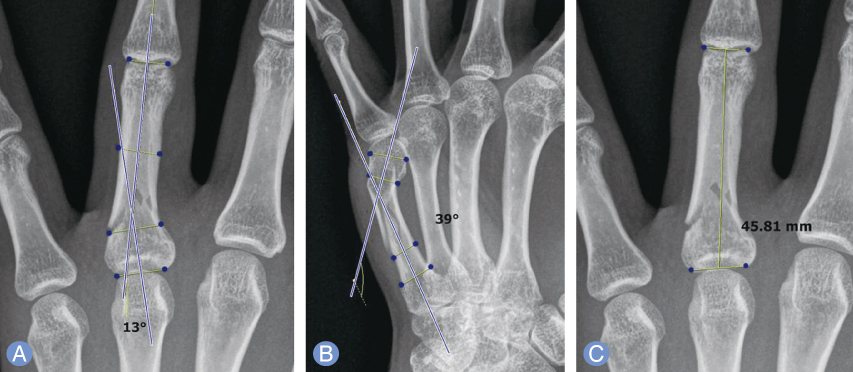
This study included 28 patients (18 male, 10 female) who had metacarpal and phalangeal fractures with rotational malalignment of finger on initial examination. Patients with combined injuries including open soft tissue damage or multiple fractures were excluded. Mean age was 36.1 years and average follow-up period was 14.6 months. Perioperative extent of rotation and correction during the follow-up, union on the radiographs, Range of motion, disability of the arm, shoulder and hand (DASH) score, and pinch power at the last follow-up were evaluated.
RESULTS
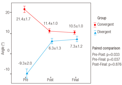
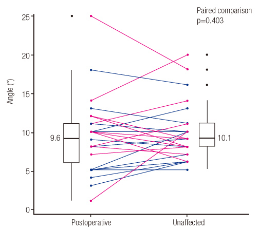
Average corrected angulation of rotation was 11.9° and no patient showed scissoring appearance of fingers at the last follow-up. All patients showed solid bony union on the radiographs during the follow-up. The average of total active motion of the injured fingers were average 254°, average DASH score was 3.2 and average pinch power was 3.0 kg at the last follow-up.
CONCLUSION
Clinical and radiologically satisfactory results were obtained in all patients. Care should be taken not to overlook the rotational misalignment after fracture of the hand, and surgical treatment should be considered to ensure correct reduction and fixation.
MeSH Terms
Figure
Reference
-
1. Chung KC, Spilson SV. The frequency and epidemiology of hand and forearm fractures in the United States. J Hand Surg Am. 2001; 26:908–915.
Article2. Barton NJ. Fractures of the hand. J Bone Joint Surg Br. 1984; 66:159–167.
Article3. Balaram AK, Bednar MS. Complications after the fractures of metacarpal and phalanges. Hand Clin. 2010; 26:169–177.
Article4. Tan V, Kinchelow T, Beredjiklian PK. Variation in digital rotation and alignment in normal subjects. J Hand Surg Am. 2008; 33:873–878.
Article5. Calandruccio JH, Jobe MT. Fractures, dislocations, and ligamentous injuries. In : Campbell WC, Canale ST, Beaty JH, editors. Campbell's operative orthopaedics. Philadelphia: Mosby/Elsevier;2008. p. 3921–3980.6. Henry MH. Hand fractures and dislocations. In : Bucholz RW, Rockwood CA, Green DP, editors. Rockwood and Green's fractures in adults. 7th ed. Philadelphia: Lippincott, Williams & Wilkins;2010. p. 711–780.7. Day CS. Fractures of the metacarpals and phalanges. In : Wolfe SW, Hotchkiss RN, Pederson WC, Kozin SH, Cohen MS, Green DP, editors. Green's operative hand surgery. 7th ed. Philadelphia: Elsevier;2017. p. 231–277.8. Jupiter JB, Axelrod TS, Belsky MR. Fractures and dislocations fo the hand. In : Browner BD, Green NE, editors. Skeletal trauma: basic science, management, and reconstruction. 3rd ed. Philadelphia: Saunders;2003. p. 1153.9. Ashkenaze DM, Ruby LK. Metacarpal fractures and dislocations. Orthop Clin North Am. 1992; 23:19–33.
Article10. Henry MH. Fractures of the proximal phalanx and metacarpals in the hand: preferred methods of stabilization. J Am Acad Orthop Surg. 2008; 16:586–595.
Article11. Lenoble E, Goutallier D. Reduction and osteosynthesis of displaced fractures of the distal third of the fifth metacarpal with central medullary bone wires. Ann Chir Main Memb Super. 1993; 12:189–195.12. Royle SG. Rotational deformity following metacarpal fracture. J Hand Surg Br. 1990; 15:124–125.
Article13. Opgrande JD, Westphal SA. Fractures of the hand. Orthop Clin North Am. 1983; 14:779–792.
Article
- Full Text Links
- Actions
-
Cited
- CITED
-
- Close
- Share
- Similar articles
-
- Miniplate and Miniscrew Fixation for the Metacarpal and Phalangeal Fractures
- Snapping Metacarpo-Phalangeal Joint after Depressed Fracture of Metacarpal Neck: Case Report
- Usefulness of Miniplate Fixation for the Fractures of Metacarpal and Phalangeal Bones of the Hand
- Treatment of rotational malalignment after interlocking intramedullary nailing of femur : Report of a case
- The Results of Miniplate Fixation for the Fractures of Metacarpal and Phalangeal Bones of the Hand


