Morphology of cat vomeronasal organ non-sensory epithelium during postnatal development
- Affiliations
-
- 1Department of Histology, Faculty of Medicine, Assiut University, Assiut, Egypt. selgar1@hotmail.com
- KMID: 2390422
- DOI: http://doi.org/10.5115/acb.2017.50.1.17
Abstract
- The vomeronasal organ has an important role in mammal's social and sexual behaviours. In addition, it mediates defensive behavior through detection of protein pheromone homologues. In this work, a detailed morphological description of the postnatal development of the non-sensory epithelium (NSE) lining the vomeronasal duct (VND) of the female cat is provided using various histological techniques. The study focused on newborn, 2 weeks, 4 weeks, and 8 weeks of postnatal ages using four animals for each age. We report here for the first time that three types of NSE line the rostral segment of the VND; nonkeratinized stratified squamous epithelium, stratified cuboidal epithelium, and ciliated pseudo stratified columnar ciliated epithelium with goblet cells and that the VND undergoes 90° a change in its its axis from the vertical position caudally to the horizontal position rostral. The NSE which lines the lateral side of the VND middle segment is consists of cliated pseudostratified columnar epithelium without goblet cells. In addition to basal cells, the NSE contains ciliated and three types of nonciliated columnar epithelial cells (dark, light, and unstained). Mitotic figures were observed only in the basal cells layer during the first 2 weeks of postnatal development. Intraepithelial invading inflammatory cells were uncommon. Scanning electron microscopy revealed unevenly distributed long cilia intermingled with microvillar processes and intervening short microvillar processes. These projecting cilia and microvilli revealed a gradual increase in their height during development toward maturity.
Keyword
MeSH Terms
Figure
-
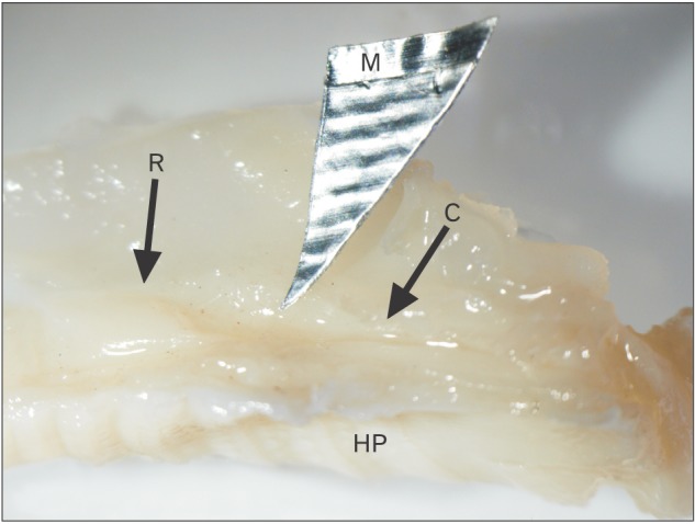
Fig. 1 Lateral view of new born cat nasal cavity showing the vomeronasal organ located at the base of the nasal septum above the hard palate (HP). It is formed of three segments; rostral (R), middle (M), and caudal (C) segments. Magnification: ×4.
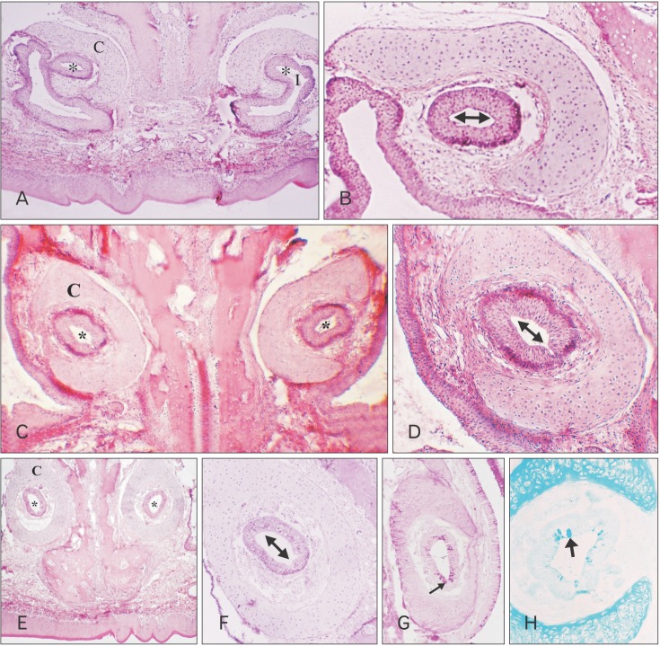
Fig. 2 New born cat. (A) The most anterior part of the vomeronasal organ (VNO) rostral segment (RS). Note the transverse direction of the lumen of the vomeronasal duct (VND) (*) which is connected to the incisive duct (I). C, cartilage. (B) Higher magnification of the previous figure showing the VND with a transversely directed lumen (↔ lined with nonkeratinized stratified squamous epithelium). (C) The middle part of the VNO RS, showing the obliquely direction of the lumen (*) of the VND. (D) Higher magnification of the previous figure showing the ciliated stratified columnar epithelium lining the middle part of the rostral segment of the VND with an obliquely directed lumen (↔). (E) The posterior part of the VNO RS, showing the vertical direction of the lumen (*) of the VND. (F) Higher magnification of the previous figure showing the pseudostratified columnar ciliated epithelium lining the posterior part of the rostral segment of the VND. (G) In the posterior part of the RS of the VNO, showing periodic acid Schiff (PAS) positive reaction in VND epithelium (arrow). (H) Transverse paraffin section in the posterior part of the RS of the VNO, showing Alcian blue (AB) positive reaction in VND epithelium (arrow). A–F, H&E staining; G, PAS staining; H, AB staining. Magnification: A, C, E, ×40; B, D, F, G, ×100; H, ×200.
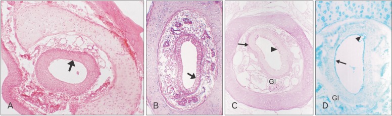
Fig. 3 Eight-week-old cat. (A) The anterior part of the rostral segment (RS) of the vomeronasal organ (VNO) is lined with stratified squamous epithelium (arrow). (B) The posterior part of RS of the VNO is lined with pseudostratified columnar ciliated epithelium with goblet cells (arrow). (C) The posterior part of the RS of the VNO vomeronasal duct (arrow) shows periodic acid Schiff (PAS) positive reaction with a few goblet cells (arrowhead) in the lining epithelium. The vomeronasal glands (Gl) in the lamina propria reveal PAS positive reaction. (D) The posterior part of the RS of the VNO shows positive Alcian blue (AB) staining of the surface epithelium (arrow) with some goblet cells (arrowhead, whereas the vomeronasal glands in the lamina propria are AB negative). A and B, H&E staining; C, PAS staining; D, AB staining. Magnification: A–C, ×100; D, ×200.
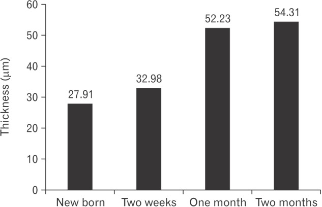
Fig. 4 Thickness of non-sensory epithelium.
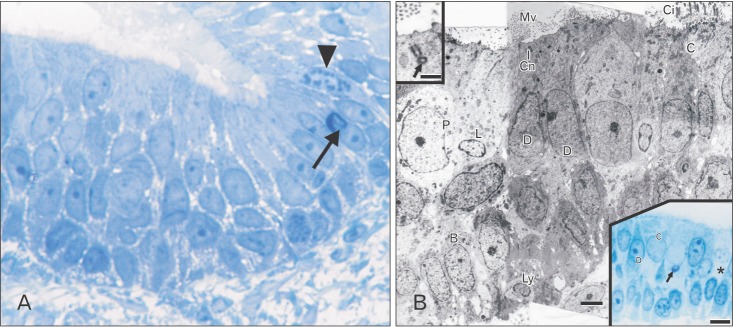
Fig. 5 New born cat. (A) Semithin section in the middle segment of vomeronasal duct at the junction between the lining vomero-sensory epithelium and non-sensory epithelium showing apical mitotic (arrowhead), and apoptotic figures (arrow) with marginal darkening of the nucleus. (B) Transmission electron microscope showing the different types of cells; ciliated cells (C) with ciliary processes (Ci), dark (D), light (L), and pale (P) non-ciliated columnar cells with microvillar proceeses (Mv) and basal cells (B). Ly, lymphocyte; Cn, centrosome. Upper inset: higher magnification for the apical present in the apical part of the cytoplasm of a dark nonciliated columnar cell. Lower inset: semithin section showing a pale nonciliated columnar cell (*). Ciliated cell (C) contains a cytoplasmic metachromatically stained secretory material (arrow). A and lower inset in B, toluidine blue staining. Magnification: A, ×1,000. Scale bars=4 µm (B), 1 µm (upper inset in B), 10 µm (lower inset in B).
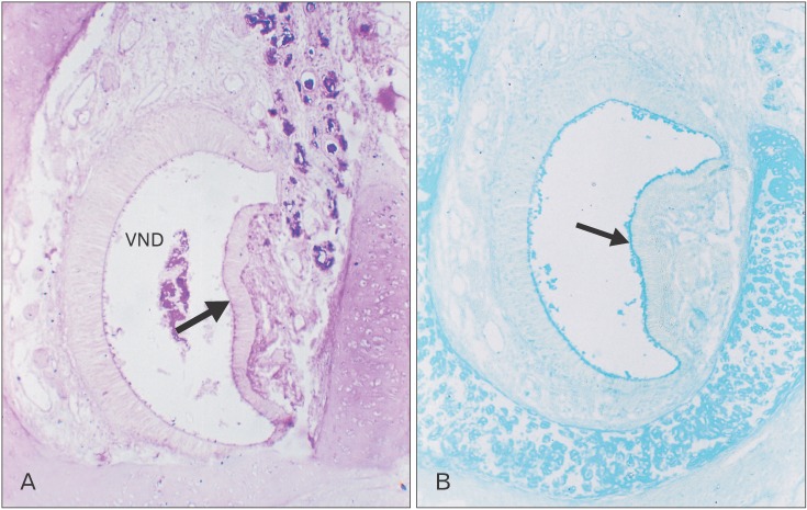
Fig. 6 Two-week-old cat. (A) Periodic acid Schiff positive reaction of the epithelial surface (arrow). (B) Alcian blue positive reaction of the epithelial surface (arrow). VND, vomeronasal duct. Magnification: A and B, ×1,000.
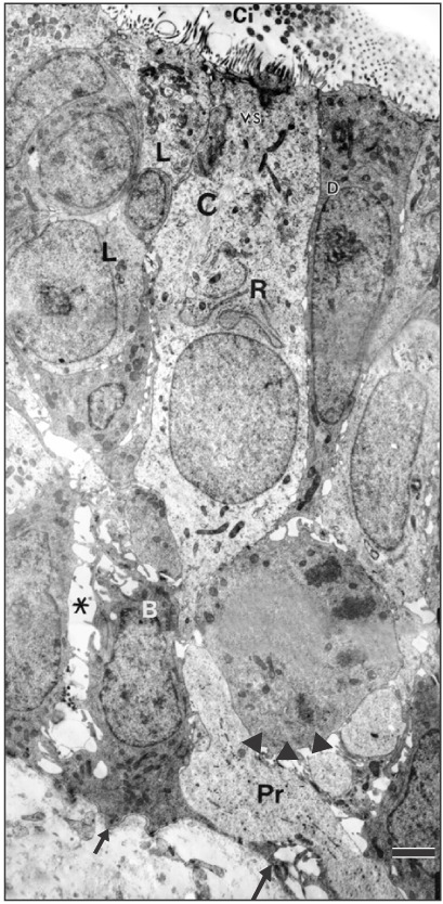
Fig. 7 Two-week-old cat. Transmission electron micrograph showing a ciliated cell (C) with a mild increase in the apical cytoplasmic organelles and the basal process (Pr). The dark (D) and light (L) nonciliated cells, exhibit an increase in supranuclear mitochondria. The basal cells (B) reveal intercellular spaces (*), lateral, and basal (arrows) processes. Note the mitotic figure present among the basal cells (arrowheads). Ci, cilia; VS, vesicles; R, rough endoplasmic reticulum. Scale bar=2.5 µm.
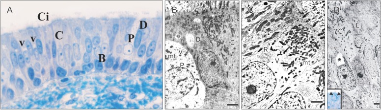
Fig. 8 Four-week-old cat. (A) Semithin section showing ciliated cells (C) with supranuclear vacuoles (V), nonciliated light pale (P), light, and dark (D) columnar cells and basal (B) cells. Note the paucity of invading cells. Ci, cilia. (B) Transmission electron microscope (TEM) showing a pale nonciliated columnar cell (P) containing vesicles (V), mitochondria (mt) and a few free ribosomes. C, ciliated; D, dark. (C) TEM showing the apical part of a ciliated cell with supranuclear vacuole (V), numerous stacks of rough endoplasmic reticulum (R) and mitochondria (mt) aggregating under the cell surface. The adjacent non-ciliated dark columnar cell contains evenly distributed mitochondria (mt). (D) TEM showing the presence of non-membrane bound irregular large supranuclear cytoplasmic vacuoles in a ciliated cell. Inset: semithin section showing metachromatically stained cytoplasmic secretory material in a ciliated cell (arrowheads). A and inset in D, toluidine blue staining. Magnification: A, ×1,000. Scale bars=4 µm (B), 2.5 µm (C), 3 µm (D), 10 µm (inset in D).
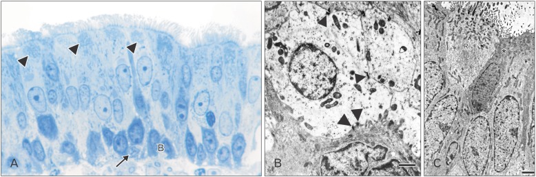
Fig. 9 Eight-week-old cat. (A) Semithin section in the non-sensory epithelium (NSE) showing an increase in the cytoplasmic cellular organelles reflected by the presence of granulations in the apical parts of the ciliated cells cytoplasm (arrowheads). An invading cell (arrow). B, basal. (B) Transmission electron microscope (TEM) for a nonciliated pale columnar cell revealing numerous mitochondria and some coated vesicles. Numerous desmosomal junctions attach the cell to adjacent cells (arrowheads). (C) TEM showing the apical part of a ciliated cell containing aggregations of mitochondria and areas of free and attached ribosomes (R). Some microvillar processes arise indirectly from the cytoplasmic extensions (arrow) of the cell. Asterisk indicates dark non-ciliated columnar cell. mv, microvilli; R, free and attached ribosomes. A, toluidine blue staining. Magnification: A, ×1,000. Scale bars=3 µm (B), 2.5 µm (C).
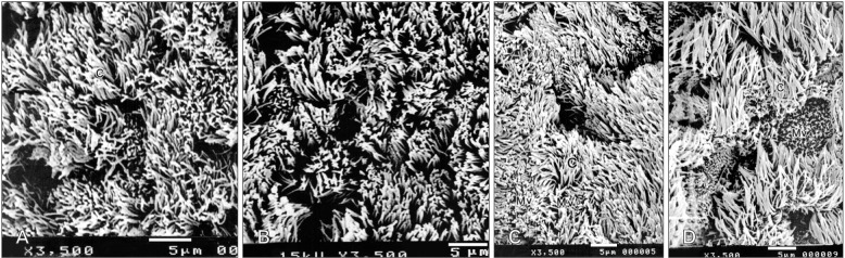
Fig. 10 Scanning electron microscope. (A) New born cat: The surface is mainly covered by ciliary (C) and microvillar processes of the same height with intervening few areas of short microvilli. (B) Two-week-old cat: note the mild increase in the height of surface processes. (C) One-month-old cat: a marked increase in the height of the surface processes has occurred. C, cilia; Mv, microvilli. (D) Two-month-old cat: a mild increase in the height of the surface processes has occurred. Scale bars=5 µm (A–D).
Reference
-
1. Brennan PA. The vomeronasal system. Cell Mol Life Sci. 2001; 58:546–555. PMID: 11361090.2. Keverne EB. Pheromones, vomeronasal function, and gender-specific behavior. Cell. 2002; 108:735–738. PMID: 11955427.3. Meredith M. Chronic recording of vomeronasal pump activation in awake behaving hamsters. Physiol Behav. 1994; 56:345–354. PMID: 7938248.4. Garrosa M, Coca S, Mora OA. Histological development of the vomeronasal complex in the pre- and postnatal rat. Acta Otolaryngol. 1986; 102:291–301. PMID: 3776523.5. Garrosa M, Coca S. Postnatal development of the vomeronasal epithelium in the rat: an ultrastructural study. J Morphol. 1991; 208:257–269. PMID: 1920442.6. Garrosa M, Iñiguez C, Fernandez JM, Gayoso MJ. Developmental stages of the vomeronasal organ in the rat: a light and electron microscopic study. J Hirnforsch. 1992; 33:123–132. PMID: 1447518.7. Weiler E. Postnatal development of the rat vomeronasal organ. Chem Senses. 2005; 30(Suppl 1):i127–i128. PMID: 15738073.8. Nakano T, Hasegawa K, Tomatsu M, Muto H. Postnatal transformation and location of mitoses in the epithelium lining the mouse vomeronasal organ. Okajimas Folia Anat Jpn. 1990; 67:81–88. PMID: 2216316.9. Elgayar SA, Eltony SA, Othman MA. Morphology of non-sensory epithelium during post-natal development of the rabbit vomeronasal organ. Anat Histol Embryol. 2014; 43:282–293. PMID: 23931650.10. Taniguchi K, Taniguchi K, Mochizuki K. Developmental studies on the vomeronasal organ in the golden hamster. Nihon Juigaku Zasshi. 1982; 44:709–716. PMID: 7161988.11. Taniguchi K, Taniguchi K. Embryonic and postnatal differentiation of olfactory epithelium and vomeronasal organ in the Syrian hamster. J Vet Med Sci. 2008; 70:57–64. PMID: 18250573.12. Irwin PJ. Companion animal parasitology: a clinical perspective. Int J Parasitol. 2002; 32:581–593. PMID: 11943231.13. Salazar I, Sanchez Quinteiro P, Cifuentes JM, Garcia Caballero T. The vomeronasal organ of the cat. J Anat. 1996; 188(Pt 2):445–454. PMID: 8621344.14. Salazar I, Sánchez-Quinteiro P. A detailed morphological study of the vomeronasal organ and the accessory olfactory bulb of cats. Microsc Res Tech. 2011; 74:1109–1120. PMID: 21484946.15. Drury RA, Wallington EA. Carelton's histological technique. 5th ed. Oxford: Oxford University Press;1980. p. 237–239.16. Gupta PD. Ultrastructural study on semithin section. Sci Tools. 1983; 30:6–7.17. Reynolds ES. The use of lead citrate at high pH as an electron-opaque stain in electron microscopy. J Cell Biol. 1963; 17:208–212. PMID: 13986422.18. Naguro T, Breipohl W. The vomeronasal epithelia of NMRI mouse: a scanning electron-microscopic study. Cell Tissue Res. 1982; 227:519–534. PMID: 7151135.19. Meisami E, Louie J, Hudson R, Distel H. A morphometric comparison of the olfactory epithelium of newborn and weanling rabbits. Cell Tissue Res. 1990; 262:89–97. PMID: 2257619.20. Coppola DM, Budde J, Millar L. The vomeronasal duct has a protracted postnatal development in the mouse. J Morphol. 1993; 218:59–64. PMID: 8230236.21. Coppola DM, Millar LC. Stimulus access to the accessory olfactory system in the prenatal and perinatal rat. Neuroscience. 1994; 60:463–468. PMID: 8072692.22. Steinberg H. Description de l'organe de Jacobson chez un foetus de chat. Anat Anz. 1912; 42:466–472.23. Kratzing JE. The olfactory apparatus of the bandicoot (Isoodon macrourus): fine structure and presence of a septal olfactory organ. J Anat. 1978; 125:601–613. PMID: 640961.24. Adams DR, Wiekamp MD. The canine vomeronasal organ. J Anat. 1984; 138(Pt 4):771–787. PMID: 6746408.25. Ponzio R. Sistema de endomembranas. In : De Robertis ED, Hib J, Ponzio R, editors. Biologia Cellular y Molecular. Buenos Aires: El Ateneo;1996. p. 221–273.26. Ferrari CC, Aldana Marcos HJ, Carmanchahi PD, Affanni JM. Olfactory mucosa of the South American armadillo Chaetophractus villosus: an ultrastructural study. Anat Rec. 1998; 252:325–339. PMID: 9811211.
- Full Text Links
-
- Actions
-
Cited
- CITED
-
- Close
- Share
-
- Similar articles
-
- Histochemical study of lectin-binding patterns in the rat vomeronasal organ during postnatal development
- A morphological study of vomeronasal organ of Korean black goat (Capra aegagrus hircus)
- Histochemical Localization of NADPH-Diaphorase in the Rat Vomeronasal Organ
- Histochemical Characterization of the Lectin-binding Sites in the Equine Vomeronasal Organ
- Sensory Stimulation-dependent Npas4 Expression in the Olfactory Bulb during Early Postnatal Development
