Central giant cell lesion of the mandible in a 2-year old girl
- Affiliations
-
- 1Department of Oral and Maxillofacial Radiology, The Nippon Dental University Niigata Hospital, Niigata, Japan. oda@ngt.ndu.ac.jp
- 2Department of Pathology, The Nippon Dental University School of Life Dentistry at Niigata, Niigata, Japan.
- KMID: 2390086
- DOI: http://doi.org/10.5624/isd.2017.47.3.209
Abstract
- Central giant cell lesions are rare, benign, osteolytic, pseudocystic, solitary, localized lesions that are common in the skeletal structure, but less so in the maxillofacial region. Furthermore, to perform panoramic radiography and cone-beam computed tomography, it is necessary to prepare patients properly and to position their heads carefully. However, this can be difficult in pediatric patients, who may be anxious. In this report, we describe the case of a central giant cell lesion of the mandible in a 2-year-old girl that was evaluated with multidetector computed tomography.
MeSH Terms
Figure
-
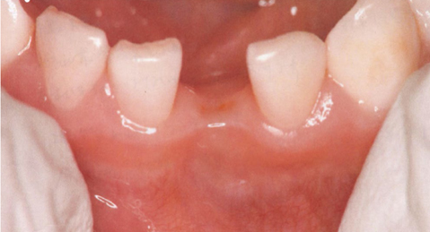
Fig. 1 A clinical intraoral photograph shows an extracted tooth wound in the central area.

Fig. 2 A periapical radiograph shows root resorption of the left mandibular lateral deciduous incisor of the mandible.
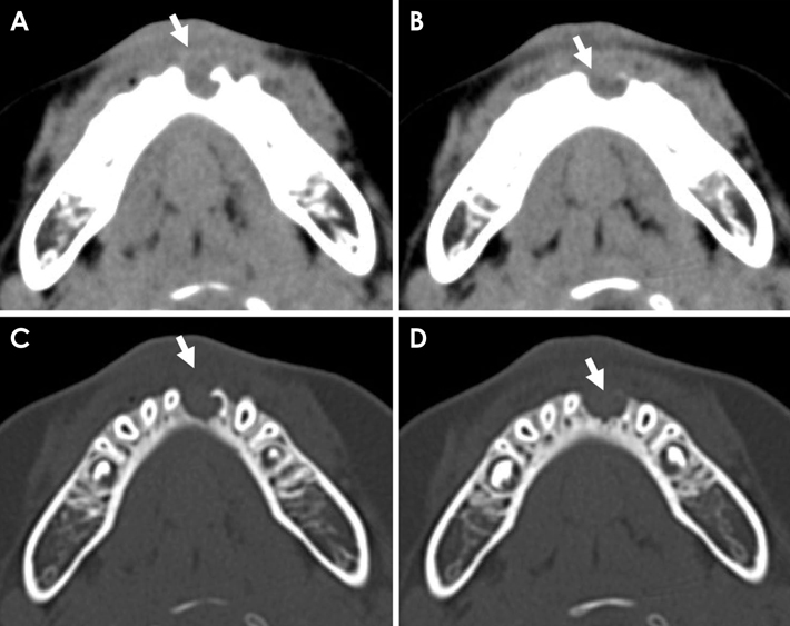
Fig. 3 Axial soft-tissue algorithm CT images (A and B) show a mass lesion along the central area (arrow). Bone-tissue algorithm CT images (C and D) show an expansile lesion with an irregular and fairly well-defined area in the central area (arrow). CT, computed tomography.
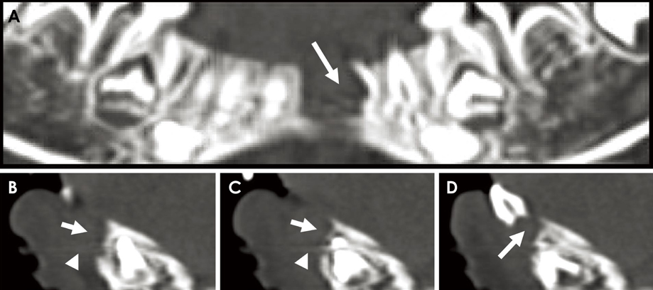
Fig. 4 Multiplanar panoramic (A) and cross-sectional (B-D) reformation images more clearly demonstrate the root resorption of the left mandibular lateral deciduous incisor of the mandible and an expansile lesion in the central area (arrow).

Fig. 5 A and B. The multinucleated osteoclast-like giant cells are embedded in a fibrous stroma comprising oval to spindle-shaped cells around the region of hemorrhage. C. Positive immunohistochemical staining for CD68.
Reference
-
1. Kulkarni D, Shetty L, Kulkarni M, Mahajan B. Extensive giant cell tumour of the mandible: a case report with review. J Maxillofac Oral Surg. 2013; 12:461–465.
Article2. Rapidis AD, Vallianatou D, Apostolidis C, Lagogiannis G. Large lytic lesion of the ascending ramus, the condyle, and the infratemporal region. J Oral Maxillofac Surg. 2004; 62:996–1001.
Article3. Rastogi S, Prashanth I, Khan SA, Trikha V, Mittal R. Giant cell tumor of bone: is curettage the answer? Indian J Orthop. 2007; 41:109–114.
Article4. Baena-Ocampo Ldel C, Ramirez-Perez E, Linares-Gonzalez LM, Delgado-Chavez R. Epidemiology of bone tumors in Mexico City: retrospective clinicopathologic study of 566 patients at a referral institution. Ann Diagn Pathol. 2009; 13:16–21.5. Jaffe HL. Giant-cell reparative granuloma, traumatic bone cyst, and fibrous (fibro-osseous) dysplasia of the jawbones. Oral Surg Oral Med Oral Pathol. 1953; 6:159–175.
Article6. Dyalram D, Aslam-Pervez N, Lubek JE. Nonodontogenic tumors of the jaws. Oral Maxillofac Surg Clin North Am. 2016; 28:59–65.
Article7. Fletcher CD, Bridge JA, Hogendoorn PC P, Mertens F. WHO classification of tumours of soft tissue and bone. 4th ed. Lyon: IARC Press;2013. p. 319–324.8. Edwards PC. Insight into the pathogenesis and nature of central giant cell lesions of the jaws. Med Oral Patol Oral Cir Bucal. 2015; 20:e196–e198.
Article9. Waldron CA, Shafer WG. The central giant cell reparative granuloma of the jaws. An analysis of 38 cases. Am J Clin Pathol. 1966; 45:437–447.
Article10. Zheng MH, Fan Y, Smith A, Wysocki S, Papadimitriou JM, Wood DJ. Gene expression of monocyte chemoattractant protein-1 in giant cell tumors of bone osteoclastoma: possible involvement in CD68+ macrophage-like cell migration. J Cell Biochem. 1998; 70:121–129.
Article11. Roux S, Amazit L, Meduri G, Guiochon-Mantel A, Milgrom E, Mariette X. RANK (receptor activator of nuclear factor kappa B) and RANK ligand are expressed in giant cell tumors of bone. Am J Clin Pathol. 2002; 117:210–216.
Article12. Lindeman JH, Hanemaaijer R, Mulder A, Dijkstra PD, Szuhai K, Bromme D, et al. Cathepsin K is the principal protease in giant cell tumor of bone. Am J Pathol. 2004; 165:593–600.
Article13. Dickson BC, Li SQ, Wunder JS, Ferguson PC, Eslami B, Werier JA, et al. Giant cell tumor of bone express p63. Mod Pathol. 2008; 21:369–375.
Article14. Lee CH, Espinosa I, Jensen KC, Subramanian S, Zhu SX, Varma S, et al. Gene expression profiling identifies p63 as a diagnostic marker for giant cell tumor of the bone. Mod Pathol. 2008; 21:531–539.
Article15. Hammas N, Laila C, Youssef AL, Hindel F, Harmouch T, Siham T, et al. Can p63 serve as a biomarker for giant cell tumor of bone? A Moroccan experience. Diagn Pathol. 2012; 7:130.
Article16. Jadu FM, Pharoah MJ, Lee L, Baker GI, Allidina A. Central giant cell granuloma of the mandibular condyle: a case report and review of the literature. Dentomaxillofac Radiol. 2011; 40:60–64.
Article17. Mohan RP, Verma S, Agarwal N, Singh U. Central giant cell granuloma: a case report. BMJ Case Rep. 2013; 2013:pii: bcr2013009903.
Article18. Park SY, Kim JY, Chang KH, Kim TJ. Giant cell tumor of the temporal bone presenting as a preauricular bulging mass. Oral Radiol. 2016; 32:56–60.
Article19. Qureshi SS, Puri A, Agarwal M, Desai S, Jambhekar N. Recurrent giant cell tumor of bone with simultaneous regional lymph node and pulmonary metastases. Skeletal Radiol. 2005; 34:225–228.
Article20. Zorlu F, Selek U, Soylemezoglu F, Oge K. Malignant giant cell tumor of the skull base originating from clivus and sphenoid bone. J Neurooncol. 2006; 76:149–152.
Article21. Patel R, Reid RR, Poon CS. Multidetector computed tomography of maxillofacial fractures: the key to high-impact radiological reporting. Semin Ultrasound CT MR. 2012; 33:410–417.
Article22. Ogura I, Sasaki Y, Kaneda T. Analysis of mandibular condylar and glenoid fossa fractures with computed tomography. Eur Radiol. 2014; 24:902–906.
Article23. Mah JK, Danforth RA, Bumann A, Hatcher D. Radiation absorbed in maxillofacial imaging with a new dental computed tomography device. Oral Surg Oral Med Oral Pathol Oral Radiol Endod. 2003; 96:508–513.
Article
- Full Text Links
-
- Actions
-
Cited
- CITED
-
- Close
- Share
-
- Similar articles
-
- A Case of a Central Giant Cell Granuloma in the Right Zygomatic Bone
- Central giant cell granuloma in mandible: report of a case
- Multiple Synchronous Central Giant Cell Granulomas of the Maxillofacial Region: A Case Report
- Central Giant Cell Granuloma of the Mandible: A Case Report
- Giant Cell Reparative Granuloma in Soft Tissue of Foot: A Case Report
