Application of a newly developed software program for image quality assessment in cone-beam computed tomography
- Affiliations
-
- 1Department of Health Technology and Biology, Federal Institute of Bahia, Salvador, BA, Brazil. marcusradiology@gmail.com
- 2Department of Interactive Processes of Organs and Systems, Institute of Health Sciences, Federal University of Bahia, Salvador, BA, Brazil.
- 3Department of Complementary Sciences, Coimbra Health School, Polytechnic Institute of Coimbra, Portugal.
- 4Department of Medical Imaging and Radiotherapy, Coimbra Health School, Polytechnic Institute of Coimbra, Portugal.
- KMID: 2389887
- DOI: http://doi.org/10.5624/isd.2017.47.2.75
Abstract
- PURPOSE
The purpose of this study was to apply a newly developed free software program, at low cost and with minimal time, to evaluate the quality of dental and maxillofacial cone-beam computed tomography (CBCT) images.
MATERIALS AND METHODS
A polymethyl methacrylate (PMMA) phantom, CQP-IFBA, was scanned in 3 CBCT units with 7 protocols. A macro program was developed, using the free software ImageJ, to automatically evaluate the image quality parameters. The image quality evaluation was based on 8 parameters: uniformity, the signal-to-noise ratio (SNR), noise, the contrast-to-noise ratio (CNR), spatial resolution, the artifact index, geometric accuracy, and low-contrast resolution.
RESULTS
The image uniformity and noise depended on the protocol that was applied. Regarding the CNR, high-density structures were more sensitive to the effect of scanning parameters. There were no significant differences between SNR and CNR in centered and peripheral objects. The geometric accuracy assessment showed that all the distance measurements were lower than the real values. Low-contrast resolution was influenced by the scanning parameters, and the 1-mm rod present in the phantom was not depicted in any of the 3 CBCT units. Smaller voxel sizes presented higher spatial resolution. There were no significant differences among the protocols regarding artifact presence.
CONCLUSION
This software package provided a fast, low-cost, and feasible method for the evaluation of image quality parameters in CBCT.
MeSH Terms
Figure
-
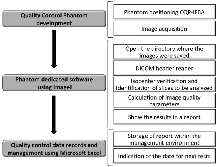
Fig. 1 Structure of study
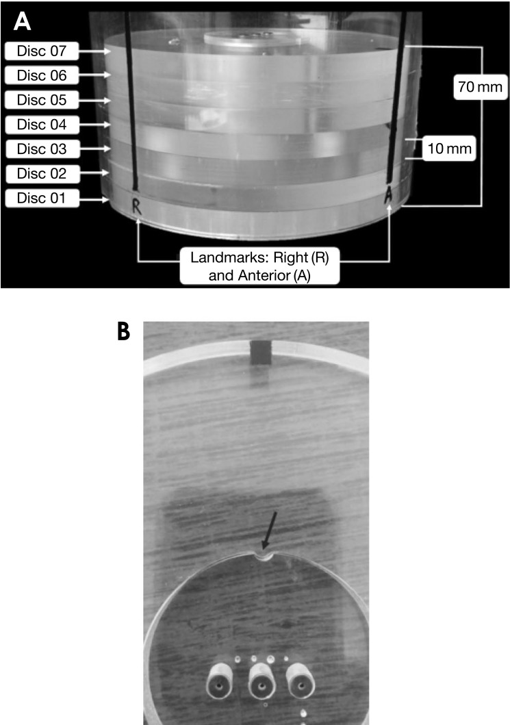
Fig. 2 Design of the phantom CQP-IFBA. A. Seven discs and location landmarks, B. docking slot inside the regular discs
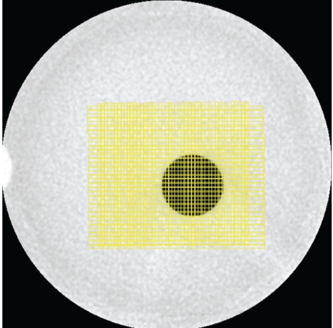
Fig. 3 Rectangular ROIs (vertical and horizontal) are drawn to identify highest standard deviation on disc 3.
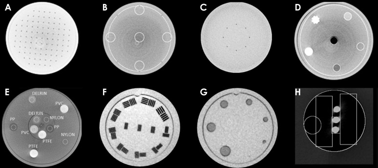
Fig. 4 Images obtained in CBCT scanners. A. Disc 1 for geometric accuracy, B. Disc 2 with ROI's positioned to evaluate the image uniformity, C. Disc 2 with a circular ROI (Ø 30 mm) in the center, D. Disc 3 with central hole containing air, E. Disc 4 used to evaluate the SNR and CNR in centered and periphery elements, F. Disc 05 with pattern bars, G. Disc 06 to evaluate the low contrast, and H. Disc 7 with three metallic rods and subtracted PMMA background
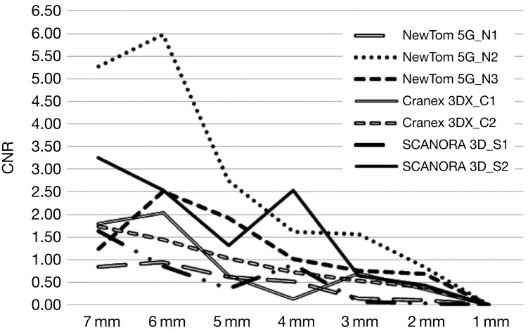
Fig. 5 Low contrast evaluation according to the protocol and contrast-to-noise ratio (CNR) in seven rods with diverse diameters
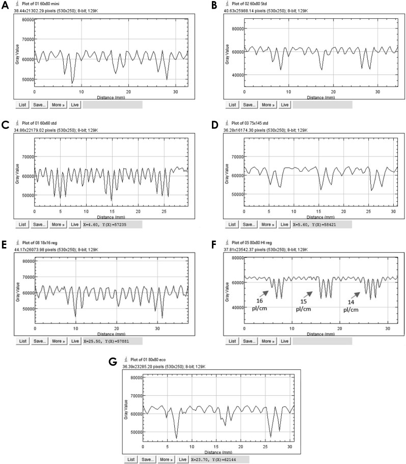
Fig. 6 Spatial resolution of CBCT units: A. Cranex 3Dx_ C1, B. Cranex 3Dx_ C2, C. Scanora 3D_ S1, D. Scanora 3D_ S2, E. NewTom 5G_ N1, F. NewTom 5G_ N2, and G. NewTom 5G_ N3
Fig. 7 Data storage system.
Reference
-
1. Miracle AC, Mukherji SK. Cone beam CT of the head and neck, part 2: clinical applications. AJNR Am J Neuroradiol. 2009; 30:1285–1292. PMID: 19461061.2. Scarfe WC, Farman AG, Sukovic P. Clinical applications of cone-beam computed tomography in dental practice. J Can Dent Assoc. 2006; 72:75–80. PMID: 16480609.3. Janner SF, Jeger FB, Lussi A, Bornstein MM. Precision of endodontic working length measurements: a pilot investigation comparing cone-beam computed tomography scanning with standard measurement techniques. J Endod. 2011; 37:1046–1051. PMID: 21763892.
Article4. Plachtovics M, Goczan J, Nagy K. The effect of calibration and detector temperature on the reconstructed cone beam computed tomography image quality: a study for the workflow of the iCAT Classic equipment. Oral Surg Oral Med Oral Pathol Oral Radiol. 2015; 119:473–480. PMID: 25687195.5. Ludlow JB, Davies-Ludlow LE, Brooks SL, Howerton WB. Dosimetry of 3 CBCT devices for oral and maxillofacial radiology: CB Mercuray, NewTom 3G and i-CAT. Dentomaxillofac Radiol. 2006; 35:219–226. PMID: 16798915.
Article6. Palomo JM, Rao PS, Hans MG. Influence of CBCT exposure conditions on radiation dose. Oral Surg Oral Med Oral Pathol Oral Radiol Endod. 2008; 105:773–782. PMID: 18424119.
Article7. Güldner C, Ningo A, Voigt J, Diogo I, Heinrichs J, Weber R, et al. Potential of dosage reduction in cone-beam-computed tomography (CBCT) for radiological diagnostics of the paranasal sinuses. Eur Arch Otorhinolaryngol. 2013; 270:1307–1315. PMID: 22986413.
Article8. Tyndall DA, Rathore S. Cone-beam CT diagnostic applications: caries, periodontal bone assessment, and endodontic applications. Dent Clin North Am. 2008; 52:825–841. PMID: 18805231.
Article9. SEDENTEXCT Guideline Development Panel. Cone beam CT for dental and maxillofacial radiology. Evidence based guidelines. Luxembourg: European Commission Directorate-General for Energy;2012.10. European Commission. Radiation Protection 136. European guidelines on radiation protection in dental radiology. Luxembourg: Office for Official Publications of the European Commission;2004.11. Bamba J, Araki K, Endo A, Okano T. Image quality assessment of three cone beam CT machines using the SEDENTEXCT CT phantom. Dentomaxillofac Radiol. 2013; 42:20120445. PMID: 23956235.
Article12. Carter L, Farman AG, Geist J, Scarfe WC, Angelopoulos C, Nair MK, et al. American Academy of Oral and Maxillofacial Radiology executive opinion statement on performing and interpreting diagnostic cone beam computed tomography. Oral Surg Oral Med Oral Pathol Oral Radiol Endod. 2008; 106:561–562. PMID: 18928899.
Article13. Torgersen GR, Hol C, Moystad A, Hellen-Halme K, Nilsson M. A phantom for simplified image quality control of dental cone beam computed tomography units. Oral Surg Oral Med Oral Pathol Oral Radiol. 2014; 118:603–611. PMID: 25442498.
Article14. Steiding C, Kolditz D, Kalender WA. A quality assurance framework for the fully automated and objective evaluation of image quality in cone-beam computed tomography. Med Phys. 2014; 41:031901. PMID: 24593719.
Article15. Pauwels R, Stamatakis H, Manousaridis G, Walker A, Michielsen K, Bosmans H, et al. Development and applicability of a quality control phantom for dental cone-beam CT. J Appl Clin Med Phys. 2011; 12:3478. PMID: 22089004.
Article16. Pauwels R, Beinsberger J, Stamatakis H, Tsiklakis K, Walker A, Bosmans H, et al. Comparison of spatial and contrast resolution for cone-beam computed tomography scanners. Oral Surg Oral Med Oral Pathol Oral Radiol. 2012; 114:127–135. PMID: 22727102.
Article17. Suomalainen A, Kiljunen T, Käser Y, Peltola J, Kortesniemi M. Dosimetry and image quality of four dental cone beam computed tomography scanners compared with multislice computed tomography scanners. Dentomaxillofac Radiol. 2009; 38:367–378. PMID: 19700530.
Article18. Loubele M, Jacobs R, Maes F, Denis K, White S, Coudyzer W, et al. Image quality vs radiation dose of four cone beam computed tomography scanners. Dentomaxillofac Radiol. 2008; 37:309–318. PMID: 18757715.19. Donini B, Rivetti S, Lanconelli N, Bertolini M. Free software for performing physical analysis of systems for digital radiography and mammography. Med Phys. 2014; 41:051903. PMID: 24784382.
Article20. Reeves TE, Mah P, McDavid WD. Deriving Hounsfield units using grey levels in cone beam CT: a clinical application. Dentomaxillofac Radiol. 2012; 41:500–508. PMID: 22752324.
Article21. Bryant JA, Drage NA, Richmond S. Study of the scan uniformity from an i-CAT cone beam computed tomography dental imaging system. Dentomaxillofac Radiol. 2008; 37:365–374. PMID: 18812597.
Article22. Lukat TD, Perschbacher SE, Pharoah MJ, Lam EW. The effects of voxel size on cone beam computed tomography images of the temporomandibular joints. Oral Surg Oral Med Oral Pathol Oral Radiol. 2015; 119:229–237. PMID: 25577416.
Article23. Pauwels R, Silkosessak O, Jacobs R, Bogaerts R, Bosmans H, Panmekiate S. A pragmatic approach to determine the optimal kVp in cone beam CT: balancing contrast-to-noise ratio and radiation dose. Dentomaxillofac Radiol. 2014; 43:20140059. PMID: 24708447.
Article24. Bechara B, Moore W, McMahan C, Noujeim M. Metal artefact reduction with cone beam CT: an in vitro study. Dentomaxillofac Radiol. 2012; 41:248–253. PMID: 22241878.25. Bechara B, Alex McMahan C, Moore WS, Noujeim M, Teixeira FB, Geha H. Cone beam CT scans with and without artefact reduction in root fracture detection of endodontically treated teeth. Dentomaxillofac Radiol. 2013; 42:20120245. PMID: 23520395.
Article26. Bechara B, McMahan C, Geha H, Noujeim M. Evaluation of a cone beam CT artefact reduction algorithm. Dentomaxillofac Radiol. 2012; 41:422–428. PMID: 22362221.
Article27. Brullmann D, Schulze RK. Spatial resolution in CBCT machines for dental/maxillofacial applications-what do we know today? Dentomaxillofac Radiol. 2015; 44:20140204. PMID: 25168812.28. Suomalainen A, Vehmas T, Kortesniemi M, Robinson S, Peltola J. Accuracy of linear measurements using dental cone beam and conventional multislice computed tomography. Dentomaxillofac Radiol. 2008; 37:10–17. PMID: 18195249.
Article
- Full Text Links
-
- Actions
-
Cited
- CITED
-
- Close
- Share
-
- Similar articles
-
- Three-dimensional imaging modalities in endodontics
- The accuracy of the imaging reformation of cone beam computed tomography for the assessment of bone defect healing
- Analysis of the priority of anatomic structures according to the diagnostic task in cone-beam computed tomographic images
- Detection of maxillary second molar with two palatal roots using cone beam computed tomography: a case report
- Basic principle of cone beam computed tomography
