A Case of Primary Small Bowel Melanoma Diagnosed by Single-Balloon Enteroscopy
- Affiliations
-
- 1Division of Gastroenterology, Department of Internal Medicine, Inha University School of Medicine, Incheon, Korea. bangbu@inha.ac.kr
- 2Department of Pathology, Inha University School of Medicine, Incheon, Korea.
- KMID: 2389246
- DOI: http://doi.org/10.5946/ce.2016.153
Abstract
- Although metastasis from cutaneous malignant melanoma to the small intestine is not uncommon, primary small bowel melanoma (SBM) is extremely rare. This case report describes a rare case of primary SBM, diagnosed by single-balloon enteroscopy. A 74-year-old man presented with recurrent melena. Upper endoscopy and colonoscopy were unremarkable. Abdominal computed tomography (CT) revealed an ileal mass with ileo-ileal intussusception. Subsequent single-balloon enteroscopy identified an ileal tumor, which was histologically diagnosed as melanoma. Extensive clinical examination did not reveal any primary cutaneous lesions. To the best of our knowledge, this is the first case of primary SBM in South Korea.
MeSH Terms
Figure
-
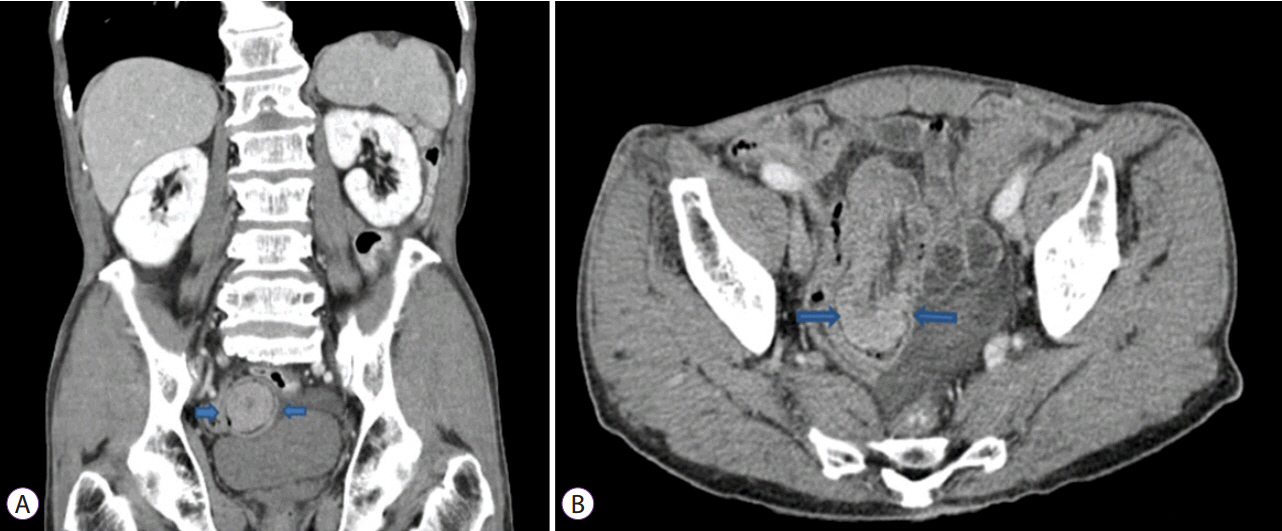
Fig. 1. Abdominal computed tomography (CT) showing 4 cm polypoid enhancing mass with ileo-ileal intussusception in the distal ileum (arrow). (A) Coronal image; (B) Axial image.

Fig. 2. Single-balloon enteroscopy showing 4 cm darkish polypoid mass obstructing the lumen approximately 60 cm proximal to the ileocecal valve.

Fig. 3. Histologic examination of endoscopic biopsy specimen showing infiltration of large atypical cells with prominent nucleoli and melanin pigment (A) and positive immunohistochemical staining for HMB-45 and S-100 (B,C). (A) Hematoxylin and eosin staining (H&E), ×40. (B) Immunohistochemical staining for HMB-45 (red color) (×200). (C) Immunohistochemical staining for S-100 protein (brown color) (×200).

Fig. 4. Gross and histologic examination of surgical specimen. (A) Gross findings showed 4 cm darkish polypoid ileal mass with regional darkish lymph node. (B) Melanin pigment is observed in the tumor cells (Hematoxylin and eosin staining [H&E], ×400). (C) Immunohistochemical staining for S-100 protein is positive (×200). (D) Immunohistochemical staining for HMB-45 is positive (×200).
Reference
-
1. Chang AE, Karnell LH, Menck HR. The American College of Surgeons Commission on Cancer and the American Cancer Society. Cancer. 1998; 83:1664–1678.2. Reintgen DS, Thompson W, Garbutt J, Seigler HF. Radiologic, endoscopic and surgical considerations of malignant melanoma metastatic to the small intestine. Curr Surg. 1984; 41:87–89.3. Timmers TK, Schadd EM, Monkelbaan JF, Meij V. Survival after resection of a primary malignant melanoma of the small intestine in a young patient: report of a case. Case Rep Gastroenterol. 2013; 7:251–260.
Article4. Hadjinicolaou AV, Hadjittofi C, Athanasopoulos PG, Shah R, Ala AA. Primary small bowel melanomas: fact or myth? Ann Transl Med. 2016; 4:113.
Article5. Mihajlovic M, Vlajkovic S, Jovanovic P, Stefanovic V. Primary mucosal melanomas: a comprehensive review. Int J Clin Exp Pathol. 2012; 5:739–753.6. Blecker D, Abraham S, Furth EE, Kochman ML. Melanoma in the gastrointestinal tract. Am J Gastroenterol. 1999; 94:3427–3433.
Article7. Das Gupta TK, Brasfield RD. Metastatic melanoma of the gastrointestinal tract. Arch Surg. 1964; 88:969–973.
Article8. Schuchter LM, Green R, Fraker D. Primary and metastatic diseases in malignant melanoma of the gastrointestinal tract. Curr Opin Oncol. 2000; 12:181–185.
Article9. Kim W, Baek JM, Suh YJ, Jeon HM, Kim JA. Ileal malignant melanoma presenting as a mass with aneurysmal dilatation: a case report. J Korean Med Sci. 2004; 19:297–301.
Article10. Amar A, Jougon J, Edouard A, Laban P, Marry JP, Hillion G. [Primary malignant melanoma of the small intestine]. Gastroenterol Clin Biol. 1992; 16:365–367.11. Krausz MM, Ariel I, Behar AJ. Primary malignant melanoma of the small intestine and the APUD cell concept. J Surg Oncol. 1978; 10:283–288.
Article12. Mishima Y. Melanocytic and nevocytic malignant melanomas. Cellular and subcellular differentiation. Cancer. 1967; 20:632–649.13. Lens M, Bataille V, Krivokapic Z. Melanoma of the small intestine. Lancet Oncol. 2009; 10:516–521.
Article14. Sachs DL, Lowe L, Chang AE, Carson E, Johnson TM. Do primary small intestinal melanomas exist? Report of a case. J Am Acad Dermatol. 1999; 41:1042–1044.
Article15. Liang KV, Sanderson SO, Nowakowski GS, Arora AS. Metastatic malignant melanoma of the gastrointestinal tract. Mayo Clin Proc. 2006; 81:511–516.
Article16. Kumari NS, Nandyala VNR, Devi KR, et al. Primary jejunal malignant melanoma presenting as intussusception: a rare case report. Int Surg J. 2014; 1:181–184.
Article17. Atmatzidis KS, Pavlidis TE, Papaziogas BT, Papaziogas TB. Primary malignant melanoma of the small intestine: report of a case. Surg Today. 2002; 32:831–833.
Article18. Butte JM, Meneses M, Waugh E, Parada H, De La Fuente H. Ileal intussusception secondary to small bowel metastases from melanoma. Am J Surg. 2009; 198:e1–e2.
Article19. Patti R, Cacciatori M, Guercio G, Territo V, Di Vita G. Intestinal melanoma: a broad spectrum of clinical presentation. Int J Surg Case Rep. 2012; 3:395–398.
Article
- Full Text Links
-
- Actions
-
Cited
- CITED
-
- Close
- Share
-
- Similar articles
-
- Diagnostic and Therapeutic Capability of Double-Balloon Enteroscopy in Clinical Practice
- Does Single Balloon Enteroscopy Have Similar Efficacy and Endoscopic Performance Compared with Double Balloon Enteroscopy?
- A Case of Primary Intestinal Lymphangiectasia Diagnosed by Double Balloon Enteroscopy
- A Case of Primary Jejunal Mucinous Adenocarcinoma Diagnosed by Single Balloon Enteroscopy
- Training in Endoscopy: Enteroscopy
