J Korean Acad Prosthodont.
2016 Oct;54(4):413-422. 10.4047/jkap.2016.54.4.413.
Prosthetic restoration of the patient with inaccurate pronunciation after prosthesis fabrication through systematic diagnosis and treatment procedure: A case report
- Affiliations
-
- 1Department of Prosthodontics, School of Dentistry, Dankook University, Cheonan, Republic of Korea. yu0324@hanmail.net
- KMID: 2388221
- DOI: http://doi.org/10.4047/jkap.2016.54.4.413
Abstract
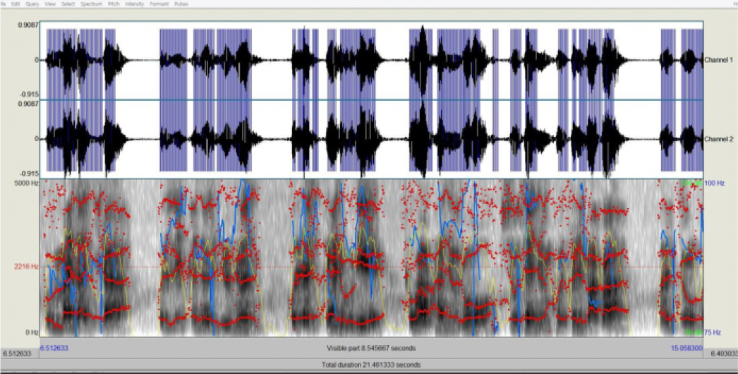
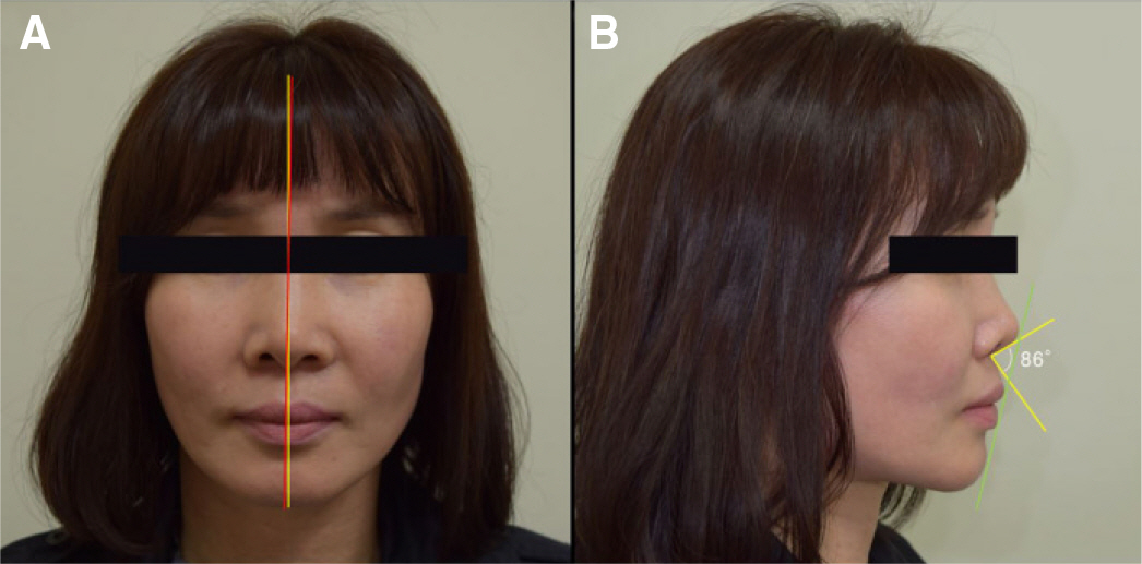
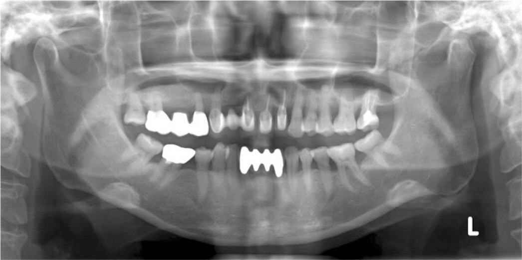
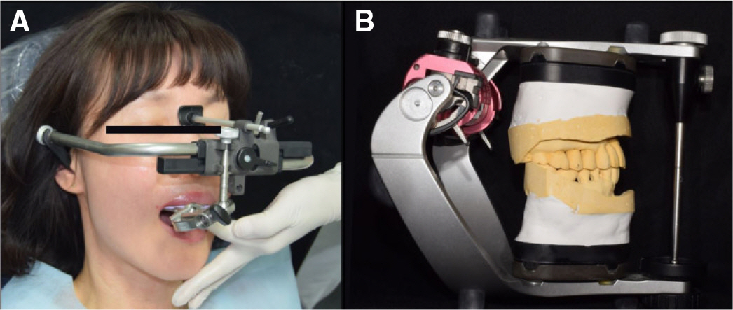
- Recently, there are cases where anterior esthetic prostheses are fabricated for better esthetics, but biologic, mechanical factors could be overlooked, too focusing on esthetic factor. This leads to changes in neutral zone, dentition, position of tongue and lips, occlusion and anterior guidance causing inaccurate pronunciation. Therefore, consideration of systematic diagnosis and treatment procedure are required. In this case, prosthesis was refabricated through a systematic diagnosis and treatment procedure using four factor (acoustic analysis, esthetic analysis, occlusion, neutral zone) for the patient who complained of inaccurate pronunciation and esthetics of the fixed prosthesis fabricated 10 years ago. Thus, by promoting functional, esthetic recovery, this case report demonstrates satisfying results to both the patient and dentist.
Figure
Cited by 1 articles
-
Maxillary anterior prosthetic treatment concerning anterior guidance of a patient who lost stable holding contact
Jong-Hoon Park, Jin-Hyun Cho
J Korean Acad Prosthodont. 2019;57(4):467-474. doi: 10.4047/jkap.2019.57.4.467.
Reference
-
1.Palmer JM. Analysis of speech in prosthodontic practice. J Prosthet Dent. 1974. 31:605–14.
Article2.Kent RA., Read C. The acoustic analysis of speech. 2nd ed.Albany: NY; Thomson Learning;2002. p. 1–60.3.Ladefoged P. A course in phonetics. 5th ed.Wadsworth: Cengage learning;2006. p. 211–36.4.Zarb GA., Bolender CL., Eckert SE., Fenton AH., Jacob RF., Mericske-Stern R. Prosthodontic treatment for edentulous patients: complete dentures and implant-supported prostheses. 12th ed.St. Louis: Mosby;2003. p. 379–88.5.Vaz Freitas S., Melo Pestana P., Almeida V., Ferreira A. Integrating voice evaluation: correlation between acoustic and audio-perceptual measures. J Voice. 2015. 29:390. .e1-7.
Article6.Ahn J., Kim G., Kim YH., Hong J. Acoustic analysis of vowel sounds before and after orthognathic surgery. J Craniomaxillofac Surg. 2015. 43:11–6.
Article7.Burris C., Vorperian HK., Fourakis M., Kent RD., Bolt DM. Quantitative and descriptive comparison of four acoustic analysis systems: vowel measurements. J Speech Lang Hear Res. 2014. 57:26–45.
Article8.Fradeani M. Evaluation of dentolabial parameters as part of a comprehensive esthetic analysis. Eur J Esthet Dent. 2006. 1:62–9.9.Calamia JR., Levine JB., Lipp M., Cisneros G., Wolff MS. Smile design and treatment planning with the help of a comprehensive esthetic evaluation form. Dent Clin North Am. 2011. 55:187–209. vii.
Article10.Mistry S. Principles of smile design-demystified. J Cosmet Dent. 2012. 28:116–24.11.Alpert RL. A method to record optimum anterior guidance for restorative dental treatment. J Prosthet Dent. 1996. 76:546–9.
Article12.Sabek M., Trévelo A. Alternative procedure for reconstructing anterior guidance using an autopolymerizing resin pattern. J Prosthet Dent. 1996. 76:550–3.
Article13.Thornton LJ. Anterior guidance: group function/canine guidance. A literature review. J Prosthet Dent. 1990. 64:479–82.
Article14.Gross MD., Cardash HS. Transferring anterior occlusal guidance to the articulator. J Prosthet Dent. 1989. 61:282–5.
Article15.Nemcovsky CE., Gross MD. Transferring provisional restorations to final master casts. J Oral Rehabil. 1994. 21:157–63.
Article16.Porwal A., Sasaki K. Current status of the neutral zone: a literature review. J Prosthet Dent. 2013. 109:129–34.
Article17.Al-Magaleh WR., Swelem AA., Shohdi SS., Mawsouf NM. Setting up of teeth in the neutral zone and its effect on speech. Saudi Dent J. 2012. 24:43–8.
Article18.Ikebe K., Okuno I., Nokubi T. Effect of adding impression material to mandibular denture space in Piezography. J Oral Rehabil. 2006. 33:409–15.
Article
- Full Text Links
- Actions
-
Cited
- CITED
-
- Close
- Share
- Similar articles
-
- CAD-CAM technique based digital diagnosis and fixed partial denture treatment on maxillary congenital missing teeth with skeletal class Ⅲ tendency patient: A case report
- All-on-6 implant fixed prosthesis restoration with fulldigital system on edentulous patient: A case report
- Maxillary anterior prosthetic treatment concerning anterior guidance of a patient who lost stable holding contact
- ‘All-on-4’ fixed implant supported prosthesis restoration using digital workflow: a case report
- Single-unit fixed restoration using the automated crown shaping artificial intelligence program

