Perinatology.
2017 Jun;28(2):29-34. 10.14734/PN.2017.28.2.29.
Clinical Features and Outcomes of Perinatally Diagnosed Meconium Peritonitis
- Affiliations
-
- 1Department of Pediatric Surgery, Keimyung University Dongsan Medical Center, Daegu, Korea. eyjung@dsmc.or.kr
- 2Department of Obsterics and Gynecology, Keimyung University Dongsan Medical Center, Daegu, Korea.
- KMID: 2386350
- DOI: http://doi.org/10.14734/PN.2017.28.2.29
Abstract
OBJECTIVE
Meconium peritonitis (MP) is defined as sterile chemical peritonitis, resulting from intrauterine bowel perforation. MP is rare but has high morbidity and mortality in neonates. We aimed to review the treatment and clinical course of MP, and to find out the possible relationship between perinatal parameters and outcomes.
METHODS
All patients diagnosed with MP between February 2006 and October 2016 were investigated retrospectively. MP was diagnosed with prenatal ultrasonography and the types of MP were identified intraoperatively. Findings of prenatal ultrasonography, gestational age, gender, birth weight, delivery type, APGAR score, clinical symptoms, causes of MP, mortality and morbidity, and hospital stay were analysed.
RESULTS
Thirteen patients were antenatally diagnosed with MP. Median gestational age was 37 weeks. All patients were diagnosed using prenatal ultrasonography. Calcification was found in 13 patients, bowel dilatation in 8, fetal ascites in 7, polyhydramnios in 6, and pseudocyst in 3. Five were females and 8 were males. Median birth weight was 2,930 g. Symptoms of abdominal distension were reported in 10 patients, bilious vomiting in 2, pneumoperitoneum in 2, and no symptoms and signs of MP in 1. One patient recovered with conservative management and the other 12 patients required surgery. All patients who underwent surgery had underlying pathologic causes; jejunoileal atresia, ileal perforation and transverse colonic perforation. Two cases of mortality occurred.
CONCLUSION
The mortality patients were haemodynamically unstable and had received preoperative pressor agents and ventilator care. More studies are needed to investigate the correlation between mortality and preoperative vital status.
Keyword
MeSH Terms
Figure
-
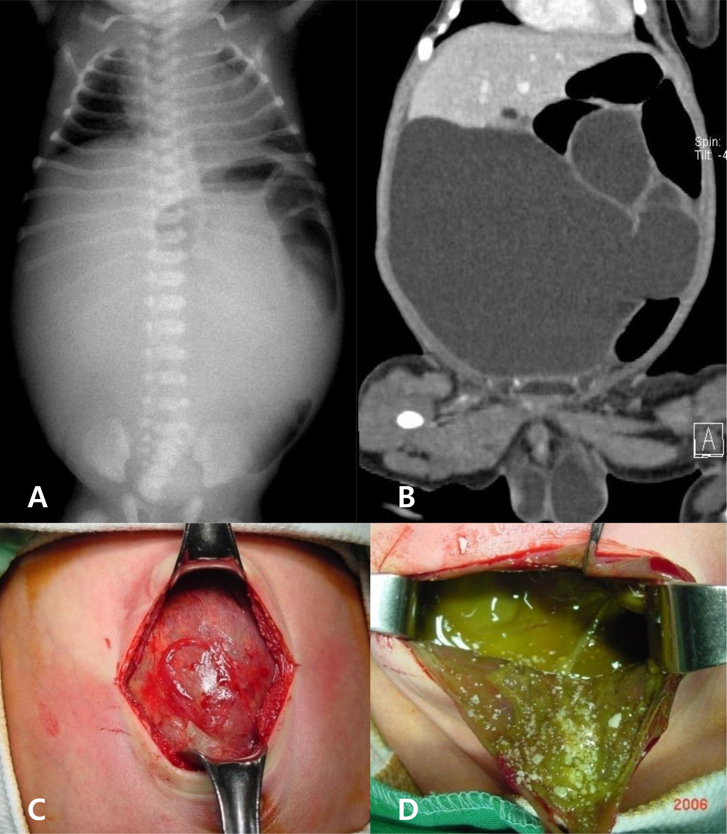
Fig. 1 Postnatal images and intraoperative findings (patient 1 in Table 2). (A) Postnatal plain X-ray; space occupying lesion in whole abdomen and bowels were positioned in left upper and lower quadrant. (B) Coronal image of computed tomography; fluid-containing cyst was seen and bowels were shifted to the left side. (C, D) Intraoperative findings; huge meconium cyst containing meconium and white calcification.
Reference
-
1). Dirkes K., Crombleholme TM., Craigo SD., Latchaw LA., Jacir NN., Harris BH, et al. The natural history of meconium peritonitis diagnosed in utero. J Pediatr Surg. 1995. 30:979–82.
Article2). Lin CH., Wu SF., Lin WC., Chen AC. Meckel's diverticulum induced intrauterine intussusception associated with ileal atresia complicated by meconium peritonitis. J Formos Med Assoc. 2007. 106:495–8.
Article3). Ekinci S., Karnak I., Akçören Z., Senocak ME. Inguinal hernia as a rare manifestation of meconium peritonitis: report of a case. Surg Today. 2008. 32:758–60.
Article4). Tongsong T., Srisupundit K., Traisrisilp K. Prenatal sonographic diagnosis of congenital varicella syndrome. J Clin Ultrasound. 2012. 40:176–8.
Article5). Foster MA., Nyberg DA., Mahony BS., Mack LA., Marks WM., Raabe RD. Meconium peritonitis: prenatal sonographic findings and their clinical significance. Radiology. 1987. 165:661–5.
Article6). Ohmichi M., Kanai H., Kanzaki T., Matumoto K., Neki R., Chiba Y, et al. Meconium peritonitis: changes in fetal C-reactive protein and CA 125 levels in relation to stage of disease. J Ultrasound Med. 1997. 16:289–92.
Article7). Martínez Ibáñez V., Boix-Ochoa J., Lloret Roca J., Ruiz H. Meconial peritonitis: conclusions based on 53 cases. Cir Pediatr. 1990. 3:80–2.8). Nam SH., Kim SC., Kim DY., Kim AR., Kim KS., Pi SY, et al. Experience with meconium peritonitis. J Pediatr Surg. 2007. 42:1822–5.
Article9). Tsai MH., Chu SM., Lien R., Huan HR., Luo CC. Clinical manifestations in infants with symptomatic meconium peritonitis. Pediatr Neonatol. 2009. 50:59–64.
Article10). Zangheri G., Andreani M., Ciriello E., Urban G., Incerti M., Vergani P. Fetal intraabdominal calcifications from meconium peritonitis: sonographic predictors of postnatal surgery. Prenat Diagn. 2007. 27:960–3.
Article11). Ping LM., Rajadurai VS., Saffari SE., Chandran S. Meconium peritonitis: correlation of antenatal diagnosis and postnatal outcome - an institutional experience over 10 years. Fetal Diagn Ther. In press. 2016.12). AboEllail MA., Tanaka H., Mori N., Tanaka A., Kubo H., Shimono R, et al. HDlive imaging of meconium peritonitis. Ultrasound Obstet Gynecol. 2015. 45:494–6.
Article13). Dewan P., Faridi MM., Singhal R., Arora SK., Rathi V., Bhatt S, et al. Meconium peritonitis presenting as abdominal calcification: three cases with different pathology. Ann Trop Paediatr. 2011. 31:163–7.
Article14). Zerhouni S., Mayer C., Skarsgard ED. Can we select fetuses with intraabdominal calcification for delivery in neonatal surgical centres? J Pediatr Surg. 2013. 48:946–50.
Article15). Saleh N., Geipel A., Gembruch U., Heep A., Heydweiller A., Bartmann P, et al. Prenatal diagnosis and postnatal management of meconium peritonitis. J Perinat Med. 2009. 37:535–8.
Article16). Eckoldt F., Heling KS., Woderich R., Kraft S., Bollmann R., Mau H. Meconium peritonitis and pseudo-cyst formation: prenatal diagnosis and postnatal course. Prenat Diagn. 2003. 23:904–8.
Article17). Kamata S., Nose K., Ishikawa S., Usui N., Sawai T., Kitayama Y, et al. Meconium peritonitis in utero. Pediatr Surg Int. 2000. 16:377–9.
Article18). Uchida K., Koike Y., Matsushita K., Nagano Y., Hashimoto K., Otake K, et al. Meconium peritonitis: prenatal diagnosis of a rare entity and postnatal management. Intractable Rare Dis Res. 2015. 4:93–7.
Article19). Casaccia G., Trucchi A., Nahom A., Aite L., Lucidi V., Giorlandino C, et al. The impact of cystic fibrosis on neonatal intestinal obstruction: the need for prenatal/neonatal screening. Pediatr Surg Int. 2003. 19:75–8.
Article20). Sato M., Hamada Y., Kohno M., Ise K., Uchida K., Ogata H, et al. Neonatal gastrointestinal perforation in Japan: a nationwide survey. Pediatr Surg. 2017. 33:33–41.
Article
