J Korean Soc Radiol.
2014 Feb;70(2):159-162. 10.3348/jksr.2014.70.2.159.
Diagnostic Usefulness of CT in Meconium Pseudocyst in a Newborn: A Case Report
- Affiliations
-
- 1Department of Radiology, Eulji University Hospital, Daejeon, Korea. kskim@eulji.ac.kr
- KMID: 1839426
- DOI: http://doi.org/10.3348/jksr.2014.70.2.159
Abstract
- A meconium pseudocyst is rare and may result in the death of the neonate. Meconium pseudocysts should be distinguished from the cystic-type meconium peritonitis because of their differences in the treatment strategy. Various studies have reported the radiographic and sonographic findings of a meconium peritonitis. However, computed tomography (CT) findings of meconium pseudocyst have been rarely reported yet. In this report we present the CT findings of a patient with a meconium pseudocyst. Such CT findings may help in the proper diagnosis and to distinguish the cystic-type meconium peritonitis from the meconium pseudocyst.
Figure
-
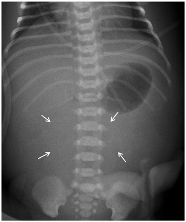
Fig. 1 Immediately after birth, the simple radiograph shows a soft tissue density mass in the mid-abdomen and egg-shell calcifications (arrows).
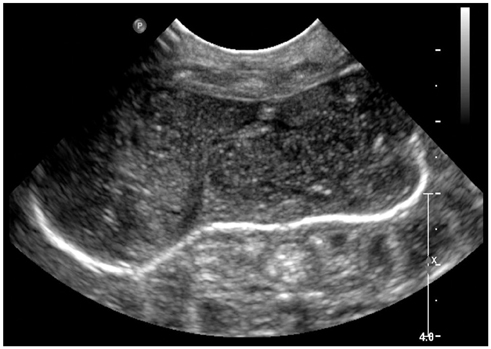
Fig. 2 Abdominal ultrasonography shows a well defined complicated cystic mass with echogenic walls separate from bowel.
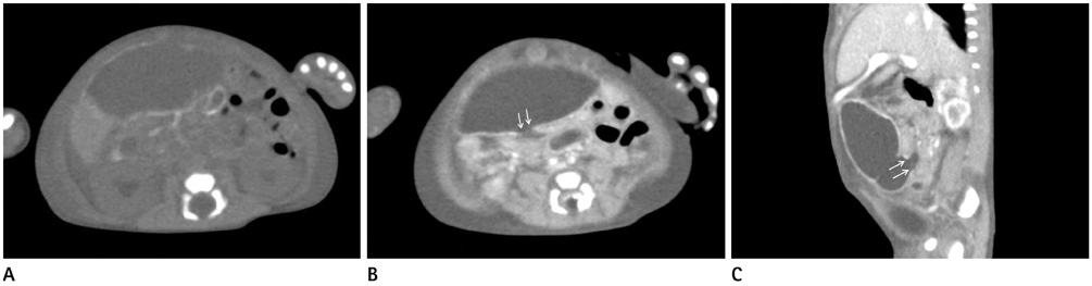
Fig. 3 A newborn with distended abdomen. A. The non-enhanced axial CT scan reveals a cystic mass with diffuse wall calcification and adjacent mesenteric calcifications. B, C. The contrast-enhanced axial (B) and sagittal CT (C) scans show a nonenhanced cystic mass with wall calcification and communication (arrows) between the cyst and adjacent dilated bowel.
Reference
-
1. Minato M, Okada T, Miyagi H, Honda S, Takazawa K, Kubota KC, et al. Meconium pseudocyst with particular pathologic findings: a case report and review of the literature. J Pediatr Surg. 2012; 47:e9–e12.2. Foster MA, Nyberg DA, Mahony BS, Mack LA, Marks WM, Raabe RD. Meconium peritonitis: prenatal sonographic findings and their clinical significance. Radiology. 1987; 165:661–665.3. Lee YC, Chen CJ. Meconium pseudocyst: a classical and successfully treated case. J Formos Med Assoc. 2009; 108:247–252.4. Shyu MK, Shih JC, Lee CN, Hwa HL, Chow SN, Hsieh FJ. Correlation of prenatal ultrasound and postnatal outcome in meconium peritonitis. Fetal Diagn Ther. 2003; 18:255–261.5. Simmons N, Shaw NJ, Mann G, Ibrahim CP. Large calcified intra-abdominal mass in a newborn. Arch Dis Child Fetal Neonatal Ed. 2009; 94:F163.6. McNamara A, Levine D. Intraabdominal fetal echogenic masses: a practical guide to diagnosis and management. Radiographics. 2005; 25:633–645.7. Salman AB, Karaoğlanoğlu N, Suma S. Abdominal, scrotal, and thoracic calcifications owing to healed meconium peritonitis. J Pediatr Surg. 1999; 34:1415–1416.8. Tanaka K, Hashizume K, Kawarasaki H, Iwanaka T, Tsuchida Y. Elective surgery for cystic meconium peritonitis: report of two cases. J Pediatr Surg. 1993; 28:960–961.9. Brenner D, Elliston C, Hall E, Berdon W. Estimated risks of radiation-induced fatal cancer from pediatric CT. AJR Am J Roentgenol. 2001; 176:289–296.
