Ann Dermatol.
2011 Nov;23(4):468-473.
IL-31 Serum Protein and Tissue mRNA Levels in Patients with Atopic Dermatitis
- Affiliations
-
- 1Department of Dermatology, Samsung Medical Center, Sungkyunkwan University School of Medicine, Seoul, Korea. junmo.yang@samsung.com
- 2Undergraduate Biological Sciences, Brown University, Providence, RI 02912, USA.
- 3Department of Statistics, Seoul National University, Seoul, Korea.
Abstract
- BACKGROUND
Severe pruritus is the primary symptom in atopic dermatitis (AD). Recently, the novel cytokine IL-31 has been implicated in the itching associated with AD.
OBJECTIVE
We performed this study to determine whether IL-31 serum levels are elevated in AD patients and to better characterize the relationship between serum IL-31 level and other established laboratory parameters.
METHODS
We recruited 55 AD patients, 34 with allergic type AD and 21 with non-allergic type AD, and 38 healthy, non-atopic controls. We checked the laboratory values, severity score, and serum IL-31 levels in all patients and controls, and IL-31 mRNA levels in lesion skin were measured in 13 subjects with AD and in four controls.
RESULTS
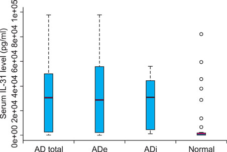
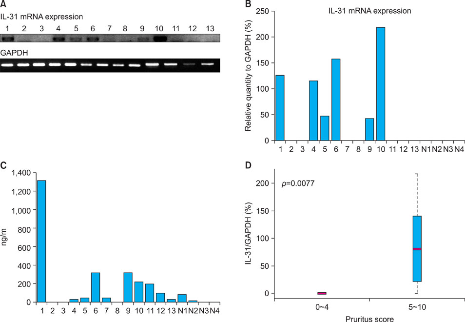
AD patients displayed significantly higher levels of serum IL-31 that were associated with serum IgE, disease severity, and subjective itch intensity. In AD patients, IL-31 mRNA levels from the lesional skin samples also correlated with serum IL-31 level.
CONCLUSION
IL-31 is likely one of the many mediators inducing inflammation and pruritus in AD. Although our limited sample size prevents us from making any definitive conclusions, our data demonstrate a strong correlation between IL-31 mRNA level and serum IL-31 protein level, which has never been reported before. Moreover, we found correlations between serum IL-31 level and serum IgE, eosinophil cationic protein, disease severity, and subject itch intensity in certain degrees in AD patients.
Keyword
MeSH Terms
Figure
Reference
-
1. Ständer S, Steinhoff M. Pathophysiology of pruritus in atopic dermatitis: an overview. Exp Dermatol. 2002. 11:12–24.
Article2. Bilsborough J, Leung DY, Maurer M, Howell M, Boguniewicz M, Yao L, et al. IL-31 is associated with cutaneous lymphocyte antigen-positive skin homing T cells in patients with atopic dermatitis. J Allergy Clin Immunol. 2006. 117:418–425.
Article3. Sonkoly E, Muller A, Lauerma AI, Pivarcsi A, Soto H, Kemeny L, et al. IL-31: a new link between T cells and pruritus in atopic skin inflammation. J Allergy Clin Immunol. 2006. 117:411–407.
Article4. Neis MM, Peters B, Dreuw A, Wenzel J, Bieber T, Mauch C, et al. Enhanced expression levels of IL-31 correlate with IL-4 and IL-13 in atopic and allergic contact dermatitis. J Allergy Clin Immunol. 2006. 118:930–937.
Article5. Dillon SR, Sprecher C, Hammond A, Bilsborough J, Rosenfeld-Franklin M, Presnell SR, et al. Interleukin 31, a cytokine produced by activated T cells, induces dermatitis in mice. Nat Immunol. 2004. 5:752–760.
Article6. Schulz F, Marenholz I, Fölster-Holst R, Chen C, Sternjak A, Baumgrass R, et al. A common haplotype of the IL-31 gene influencing gene expression is associated with nonatopic eczema. J Allergy Clin Immunol. 2007. 120:1097–1102.
Article7. Grimstad O, Sawanobori Y, Vestergaard C, Bilsborough J, Olsen UB, Grønhøj-Larsen C, et al. Anti-interleukin-31-anti-bodies ameliorate scratching behaviour in NC/Nga mice: a model of atopic dermatitis. Exp Dermatol. 2009. 18:35–43.
Article8. Jeong CW, Ahn KS, Rho NK, Park YD, Lee DY, Lee JH, et al. Differential in vivo cytokine mRNA expression in lesional skin of intrinsic vs. extrinsic atopic dermatitis patients using semiquantitative RT-PCR. Clin Exp Allergy. 2003. 33:1717–1724.
Article9. Rho NK, Kim WS, Lee DY, Lee JH, Lee ES, Yang JM. Immunophenotyping of inflammatory cells in lesional skin of the extrinsic and intrinsic types of atopic dermatitis. Br J Dermatol. 2004. 151:119–125.
Article10. Hanifin JM, Rajka G. Diagnostic feature of atopic dermatitis. Acta Derma Venerol (Stockh). 1980. 92:Suppl. 44–47.11. Severity scoring of atopic dermatitis: the SCORAD index. Consensus Report of the European Task Force on Atopic Dermatitis. Dermatology. 1993. 186:23–31.12. Leung DY, Boguniewicz M, Howell MD, Nomura I, Hamid QA. New insights into atopic dermatitis. J Clin Invest. 2004. 113:651–657.
Article13. Hamid Q, Boguniewicz M, Leung DY. Differential in situ cytokine gene expression in acute versus chronic atopic dermatitis. J Clin Invest. 1994. 94:870–876.
Article14. Ständer S, Steinhoff M, Schmelz M, Weisshaar E, Metze D, Luger T. Neurophysiology of pruritus: cutaneous elicitation of itch. Arch Dermatol. 2003. 139:1463–1470.15. Chi KH, Myers JN, Chow KC, Chan WK, Tsang YW, Chao Y, et al. Phase II trial of systemic recombinant interleukin-2 in the treatment of refractory nasopharyngeal carcinoma. Oncology. 2001. 60:110–115.
Article16. Lee CH, Chuang HY, Shih CC, Jong SB, Chang CH, Yu HS. Transepidermal water loss, serum IgE and beta-endorphin as important and independent biological markers for development of itch intensity in atopic dermatitis. Br J Dermatol. 2006. 154:1100–1107.
Article17. Chan LS, Robinson N, Xu L. Expression of interleukin-4 in the epidermis of transgenic mice results in a pruritic inflammatory skin disease: an experimental animal model to study atopic dermatitis. J Invest Dermatol. 2001. 117:977–983.
Article18. Takaoka A, Arai I, Sugimoto M, Yamaguchi A, Tanaka M, Nakaike S. Expression of IL-31 gene transcripts in NC/Nga mice with atopic dermatitis. Eur J Pharmacol. 2005. 516:180–181.
Article19. Raap U, Wichmann K, Bruder M, Stnder S, Wedi B, Kapp A, et al. Correlation of IL-31 serum levels with severity of atopic dermatitis. J Allergy Clin Immunol. 2008. 122:421–423.
Article
- Full Text Links
- Actions
-
Cited
- CITED
-
- Close
- Share
- Similar articles
-
- Eosinophil Counts in Peripheral Blood, Serum Total IgE, Eosinophil Cationic Protein, IL-4 and Soluble E-selectin in Atopic Dermatitis
- Relationship between serum interleukin-31/25-hydroxyvitamin D levels and the severity of atopic dermatitis in children
- Serum IgE Level in Patients of Atopic Dermatitis and Atopic Dermatitis with Molluscum Contagiosum
- Plasma Histamine Levels in Patients with Atopic Dermatitis
- Correlation between the Level of Urinary Leukotriene E4 and Seurm Eosinophil Cationic Protein and Clinical Severity of Atopic Dermatitis in Children


