Acute Paraplegia After Lumbar Steroid Injection in Patients With Spinal Dural Arteriovenous Fistulas: Case Reports
- Affiliations
-
- 1Department of Physical and Rehabilitation Medicine, Samsung Medical Center, Sungkyunkwan University School of Medicine, Seoul, Korea. yays.sung@samsung.com
- KMID: 2382929
- DOI: http://doi.org/10.5535/arm.2016.40.5.949
Abstract
- Spinal dural arteriovenous fistulas (SDAVFs) are the most common type of spinal vascular malformations. However, SDAVFs are still underdiagnosed entities because their clinical symptoms are usually non-specific, as they include low back pain or radiating pain to the limb. There have been several reports of acute paraplegia after lumbar epidural steroid injections in patients with SDAVFs. We present 4 patients with SDAVFs who received lumbar steroid injection. Among the 4 cases, acute paraplegia developed in 2 cases that received a larger volume of injectate than the other cases. Thus, we are suggesting that the volume of injectate may be a contributing factor for acute paraplegia after lumbar steroid injection in patients with SDAVFs.
Keyword
MeSH Terms
Figure
-

Fig. 1 Sagittal T2-weighted MRI images obtained (A) on the day after the injection and (B) one day before the injection show tortuous fluid void structures suggestive of SDAVFs (arrows). (C) Spinal angiography image shows SDAVFs supplied by the dural branch of the right T6 intercostal artery (arrow) and tortuous draining veins (arrowheads). (D) On spinal angiography conducted after glue embolization, SDAVFs had disappeared. SDAVFs, spinal dural arteriovenous fistulas.
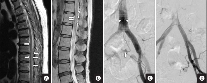
Fig. 2 (A) On MRI taken 2 days after the injection, sagittal T2-weighted image shows tortuous fluid void structures suggestive of SDAVFs (arrows). (B) On MRI taken before the injection, sagittal T2-weighted image shows tortuous fluid void structures (arrows) and increased spinal cord signal intensity of SDAVFs (arrowheads). (C) Spinal angiography image shows SDAVFs supplied by the sacral branch of the left internal iliac artery (arrow) and tortuous draining veins (arrowheads). (D) On spinal angiography conducted after glue embolization, SDAVFs had disappeared. SDAVFs, spinal dural arteriovenous fistulas.
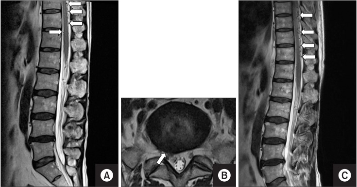
Fig. 3 (A) In the sagittal T2-weighted MRI image taken before the injection, tortuous fluid void structures suggestive of SDAVFs (arrows) are noticeable. (B) On the same MRI, axial T2-weighted image shows a herniated intervertebral disc at right the L5-S1 level (arrow). (C) On the follow-up MRI, SDAVFs persisted, and the extent of engorged perimedullary veins (arrows) is similar to that in the previous MRI. SDAVFs, spinal dural arteriovenous fistulas.
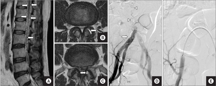
Fig. 4 (A) On the MRI taken before the injection, the sagittal T2-weighted image shows tortuous fluid void structures suggestive of SDAVFs (arrows). (B) On the same MRI, axial T2-weighted image shows degeneration of the left L3-4 facet joint (arrow). (C) Also on the same MRI, axial T2-weighted image shows degeneration of the left L4-5 facet joint (arrow). (D) Spinal angiography image shows SDAVFs supplied by the right internal iliac artery (arrow) and tortuous draining veins (arrowheads). (E) On spinal angiography conducted after glue embolization, SDAVFs and perimedullary venous engorgement had disappeared. SDAVFs, spinal dural arteriovenous fistulas.
Reference
-
1. Koch C. Spinal dural arteriovenous fistula. Curr Opin Neurol. 2006; 19:69–75. PMID: 16415680.
Article2. Kataoka H, Miyamoto S, Nagata I, Ueba T, Hashimoto N. Venous congestion is a major cause of neurological deterioration in spinal arteriovenous malformations. Neurosurgery. 2001; 48:1224–1229. PMID: 11383723.
Article3. Aminoff MJ, Logue V. The prognosis of patients with spinal vascular malformations. Brain. 1974; 97:211–218. PMID: 4434169.
Article4. Jellema K, Tijssen CC, van Gijn J. Spinal dural arteriovenous fistulas: a congestive myelopathy that initially mimics a peripheral nerve disorder. Brain. 2006; 129(Pt 12):3150–3164. PMID: 16921175.
Article5. Krings T, Geibprasert S. Spinal dural arteriovenous fistulas. AJNR Am J Neuroradiol. 2009; 30:639–648. PMID: 19213818.
Article6. Cabrera M, Paradas C, Marquez C, Gonzalez A. Acute paraparesis following intravenous steroid therapy in a case of dural spinal arteriovenous fistula. J Neurol. 2008; 255:1432–1433. PMID: 18758891.
Article7. Strowd RE, Geer C, Powers A, Abou-Zeid N. A unique presentation of a spinal dural arteriovenous fistula exacerbated by steroids. J Clin Neurosci. 2012; 19:466–468. PMID: 22249021.
Article8. Hetts SW, Narvid J, Singh T, Meagher S, Corcoran K, Higashida RT, et al. Association between lumbar epidural injection and development of acute paraparesis in patients with spinal dural arteriovenous fistulas. AJNR Am J Neuroradiol. 2007; 28:581–583. PMID: 17353341.
- Full Text Links
-
- Actions
-
Cited
- CITED
-
- Close
- Share
-
- Similar articles
-
- Endovascular Treatment of Spinal Dural and Epidural Arteriovenous Fistula as Complication of Lumbar Surgery
- Whole Lumbar Spinal Subdural Hematoma with Progressive Paraplegia after Lumbar Spinal Epidural Injection
- Spinal Arteriovenous Fistula with Progressive Paraplegia after Spinal Anaesthesia
- Fibrous Mass Complicating Epidural Steroid and Local Anesthetic Injection: A Case Report
- Surgical Treatment of Arteriovenous Malformations of the Spinal Cord
