Clin Exp Otorhinolaryngol.
2017 Jun;10(2):188-192. 10.21053/ceo.2016.00185.
Tracheobronchial Angle Measurements in Children: An Anthropometric Retrospective Study With Multislice Computed Tomography
- Affiliations
-
- 1Department of Radiology, Pamukkale University School of Medicine, Denizli, Turkey. dtherek@yahoo.com
- 2Department of Pediatric Surgery, Pamukkale University School of Medicine, Denizli, Turkey.
- KMID: 2380439
- DOI: http://doi.org/10.21053/ceo.2016.00185
Abstract
OBJECTIVES
The purpose of this study is to investigate if any change exists in the values of tracheal bifurcation angles (subcarinal angle [SCA] and interbronchial angle [IBA]), right and left bronchial angles (RBA and LBA) in different pediatric age groups.
METHODS
Chest computed tomography (CT) images of children aged 18 years and younger were reviewed retrospectively by two radiologists who were blinded to each other's measurements. One hundred and eighteen children were involved. RBA, LBA, SCA, and IBA were measured on coronal reformatted images. Subjects were classified into three groups according to their age. Measurement of IBA was done by measuring the angle between the lines drawn along the central axis of right and left main bronchi over their length. RBA and LBA were measured at the intersection points of the lines drawn along the inferior borders of the right and left main bronchi and the line passing through the longitudinal axis of trachea. Sums of RBA and LBA gave SCA. Interobserver agreement was also analyzed.
RESULTS
SCA, IBA, and RBA values were statistically significant between children of ages less than 10 years and over 10 years P<0.01). Interobserver agreement was excellent with an intraclass correlation coefficient score of 0.87 (95% confidence interval) for RBA, SCA, and IBA measurements.
CONCLUSION
We concluded that tracheal bifurcation angles are wider in children of age 10 years and younger. As age increases values of SCA, IBA, and RBA decrease.
Keyword
MeSH Terms
Figure
-
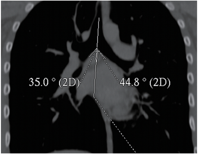
Fig. 1. Measurement of right and left bronchial angles on a coronal reformatted computed tomography image of a 16-year-old boy. Sum of these angles gives subcarinal angle. 2D, 2 dimensional.
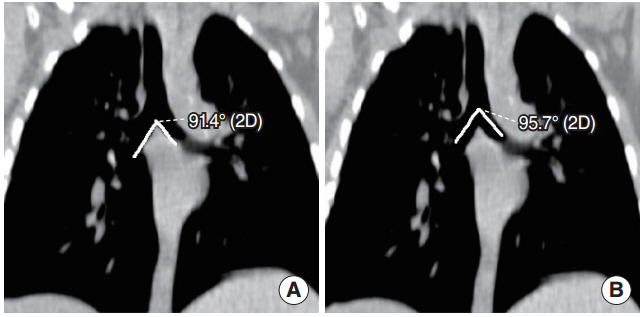
Fig. 2. Note the location of subcarinal angle (A) and interbronchial angle (B) on coronal reformatted computed tomography images of a 1-year-old boy.
Reference
-
1. Haskin PH, Goodman LR. Normal tracheal bifurcation angle: a reassessment. AJR Am J Roentgenol. 1982; Nov. 139(5):879–82.
Article2. Karabulut N. CT assessment of tracheal carinal angle and its determinants. Br J Radiol. 2005; Sep. 78(933):787–90.
Article3. Chunder R, Nandi S, Guha R, Satyanarayana N. A morphometric study of human trachea and principal bronchi in different age groups in both sexes and its clinical implications. Nepal Med Coll J. 2010; Dec. 12(4):207–14.4. Chunder R, Guha R. A morphometric study of human subcarinal angle in different age groups in both sexes and its clinical implications. Indian J Basic Appl Med Res. 2015; Mar. 4(2):424–30.5. Mi W, Zhang C, Wang H, Cao J, Li C, Yang L, et al. Measurement and analysis of the tracheobronchial tree in Chinese population using computed tomography. PLoS One. 2015; Apr. 10(4):e0123177.
Article6. Kamel KS, Lau G, Stringer MD. In vivo and in vitro morphometry of the human trachea. Clin Anat. 2009; Jul. 22(5):571–9.
Article7. Tahir N, Ramsden WH, Stringer MD. Tracheobronchial anatomy and the distribution of inhaled foreign bodies in children. Eur J Pediatr. 2009; Mar. 168(3):289–95.
Article8. Murray JG, Brown AL, Anagnostou EA, Senior R. Widening of the tracheal bifurcation on chest radiographs: value as a sign of left atrial enlargement. AJR Am J Roentgenol. 1995; May. 164(5):1089–92.
Article9. Daroszewski M, Szpinda M, Flisinski P, Szpinda A, Wozniak A, Kosinski A, et al. Tracheo-bronchial angles in the human fetus: an anatomical, digital, and statistical study. Med Sci Monit Basic Res. 2013; Jul. 19:194–200.10. Fong EW. Tracheo-bronchial angles in the human fetus: an anatomical, digital, and statistical study. In : Yamamoto L, editor. Case based pediatrics for medical students and residents. 2nd ed. Honolulu, HI: Department of Pediatrics, University of Hawaii John A. Burns School of Medicin;2002. p. 311–4.11. Itasca IL. National Safety Council: injury facts, 2001 ed. Itasca, IL: National Safety Council;2001.12. Cohen SR, Herbert WI, Lewis GB Jr, Geller KA. Foreign bodies in the airway: five-year retrospective study with special reference to management. Ann Otol Rhinol Laryngol. 1980; Sep-Oct. 89(5 Pt 1):437–42.13. Daniilidis J, Symeonidis B, Triaridis K, Kouloulas A. Foreign body in the airways: a review of 90 cases. Arch Otolaryngol. 1977; Oct. 103(10):570–3.
Article14. Van Looij MA, Rood PP, Hoeve LJ, Borgstein JA. Aspirated foreign bodies in children: why are they more commonly found on the left? Clin Otolaryngol Allied Sci. 2003; Aug. 28(4):364–7.
Article15. Baharloo F, Veyckemans F, Francis C, Biettlot MP, Rodenstein DO. Tracheobronchial foreign bodies: presentation and management in children and adults. Chest. 1999; May. 115(5):1357–62.16. Kubota Y, Toyoda Y, Nagata N, Kubota H, Sawada S, Murakawa M, et al. Tracheo-bronchial angles in infants and children. Anesthesiology. 1986; Mar. 64(3):374–6.
Article17. Adriani J, Griggs TS. An improved endotracheal tube for pediatric use. Anesthesiology. 1954; Sep. 15(5):466–70.18. Landis JR, Koch GG. The measurement of observer agreement for categorical data. Biometrics. 1977; Mar. 33(1):159–74.
Article
- Full Text Links
-
- Actions
-
Cited
- CITED
-
- Close
- Share
-
- Similar articles
-
- Helical CT of a Tracheobronchial Phantom: Angle and Optimal Window Affecting Size Measurements of ThreeDimensional Images
- Ruptured Aneurysm of the Ophthalmic Artery
- Comparison of Aneurysmal Clip-induced Artifacts in 64- and 16-row Multislice Computed Tomography Angiograms
- Comparison of Orbital Anatomy in Korean and Caucasian Patients Using Computed Tomography
- The Effect of Taste Preference on Anthropometric Measurements and Nutrient intakes in Children
