Comparison of Orbital Anatomy in Korean and Caucasian Patients Using Computed Tomography
- Affiliations
-
- 1Department of Ophthalmology, Dong-A University College of Medicine, Busan, Korea. hbahn@dau.ac.kr
- KMID: 2214543
- DOI: http://doi.org/10.3341/jkos.2015.56.9.1311
Abstract
- PURPOSE
In this study we analyzed and compared the orbital anatomy in Korean and Caucasian subjects using computed tomography (CT) measurements.
METHODS
Two observers performed a cross-sectional, retrospective analysis of 44 CT scans of subjects (22 Koreans, 22 Caucasians) with no appreciable orbital or globe disease. Ten length measurements and 3 angle measurements of various orbital aspects were obtained. Analysis was performed to determine if changes in these parameters were associated with race.
RESULTS
Anterior medial interorbital length was 24.05 +/- 2.00 mm in Korean and 21.96 +/- 1.96 mm in Caucasian subjects. Anterior vertical orbital length was 34.19 +/- 1.67 mm in Korean and 35.03 +/- 1.18 mm in Caucasian subjects. The anterior medial interorbital length and anterior vertical orbital length (p < 0.05) were significantly different. Interorbital angle was 47.69degrees +/- 1.49degrees in Korean and 46.15degrees +/- 2.19degrees in Caucasian subjects; the difference was statistically significant (p < 0.05).
CONCLUSIONS
Compared with Caucasians, the orbit of Korean subjects has a narrower orbital opening and longer interorbital distance. The present study regarding the anatomy of Korean and Caucasian orbits will assist physicians with the evaluation and treatment of orbital diseases.
Keyword
MeSH Terms
Figure
-
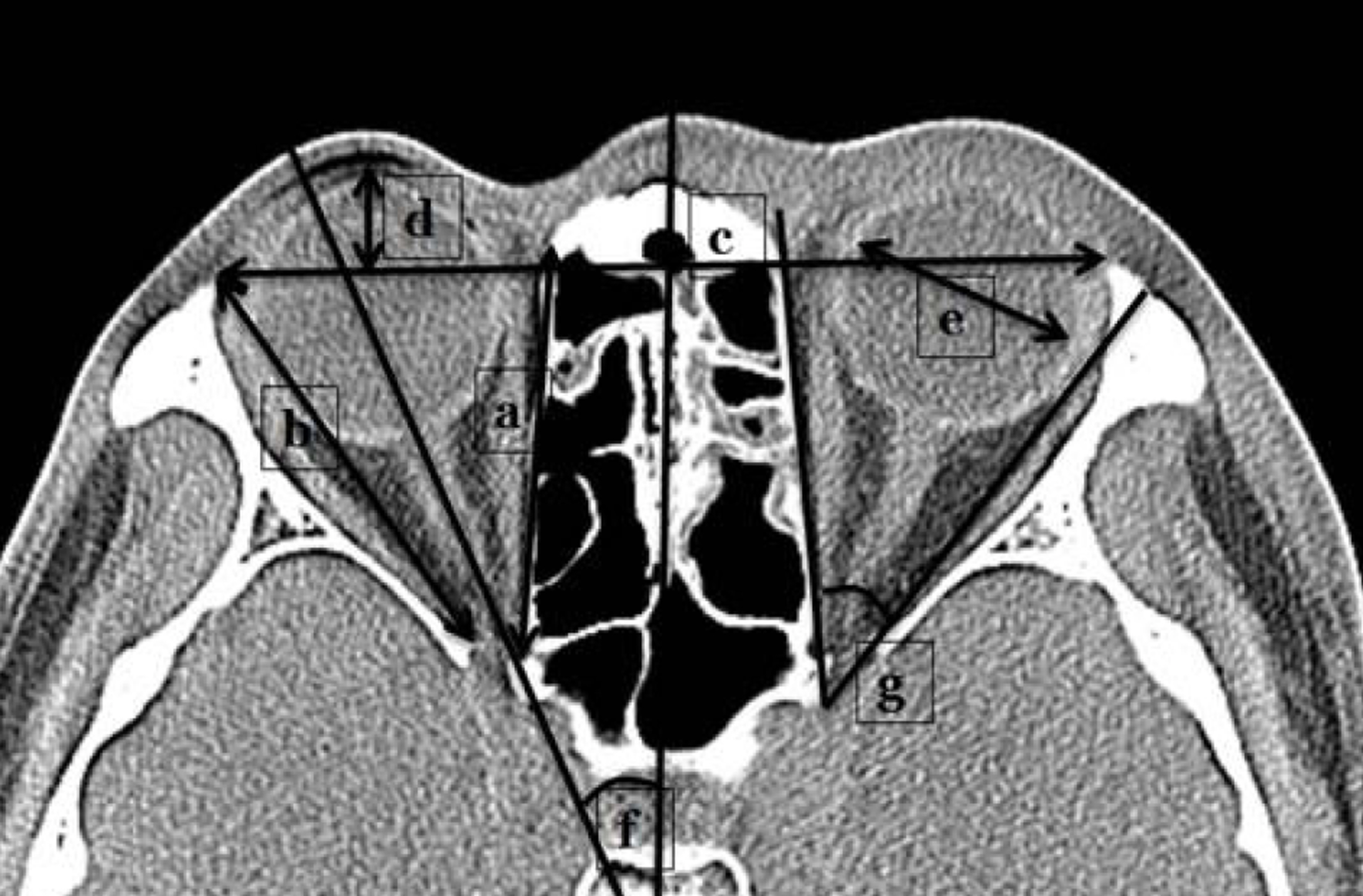
Figure 1. Axial computed tomography slice that illustrates 5 lengths and 2 orbital angle parameters recorded. ‘a’ means me-dial orbital wall length. ‘b’ means lateral orbital wall length. ‘c’ means lateral wall interorbital length. ‘d’ means globe protrusion. ‘e’ means globe diameter. ‘f’ means central orbital axis angle. ‘g’ means medial-to-lateral orbital wall angle.
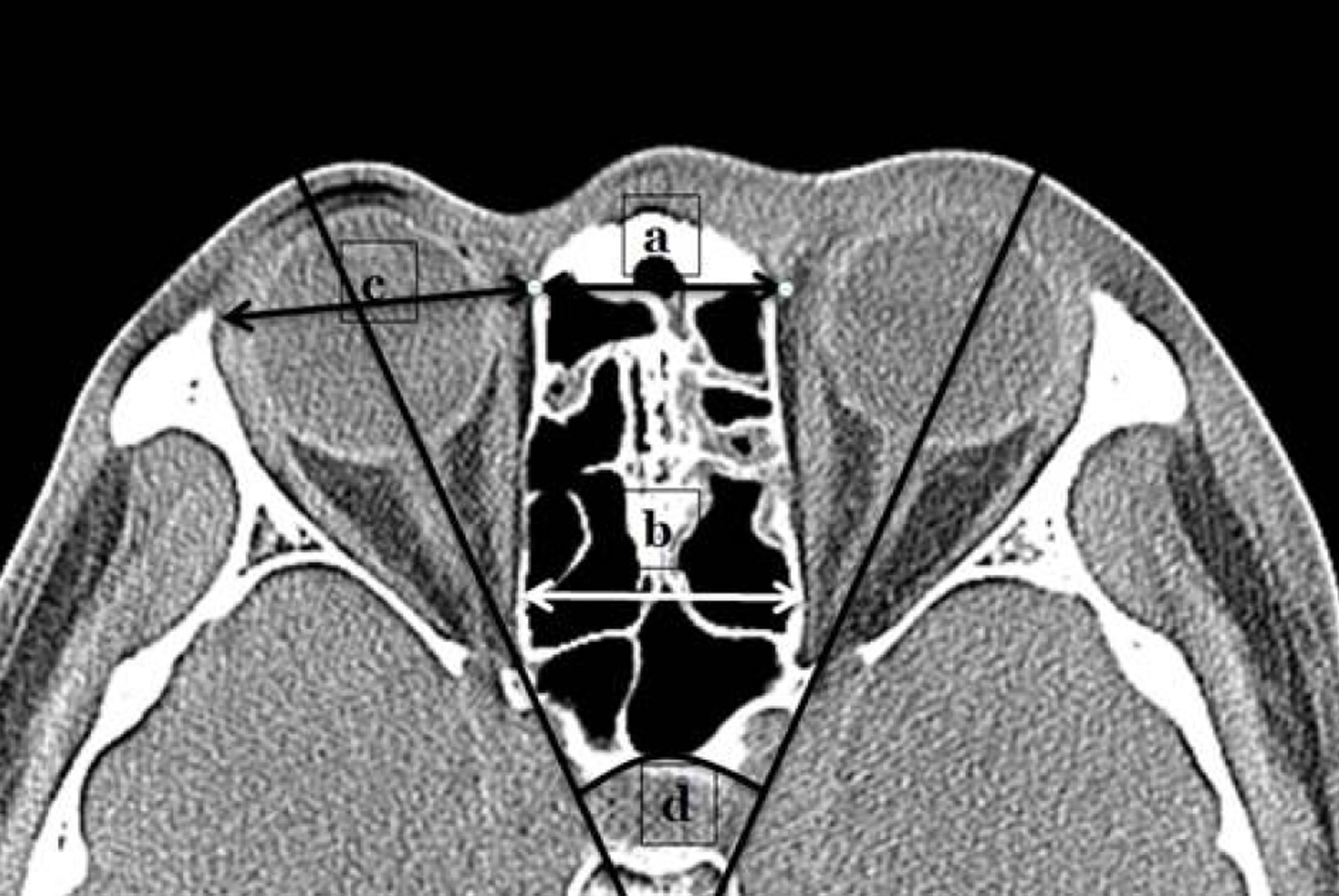
Figure 2. Axial computed tomography slice that illustrates an additional 3 lengths and 1 orbital angle parameters. ‘a’ means anterior medial interorbital length. ‘b’ means maximal medial interorbital length. ‘c’ means maximal horizontal orbital length. ‘d’ means interorbital angle.
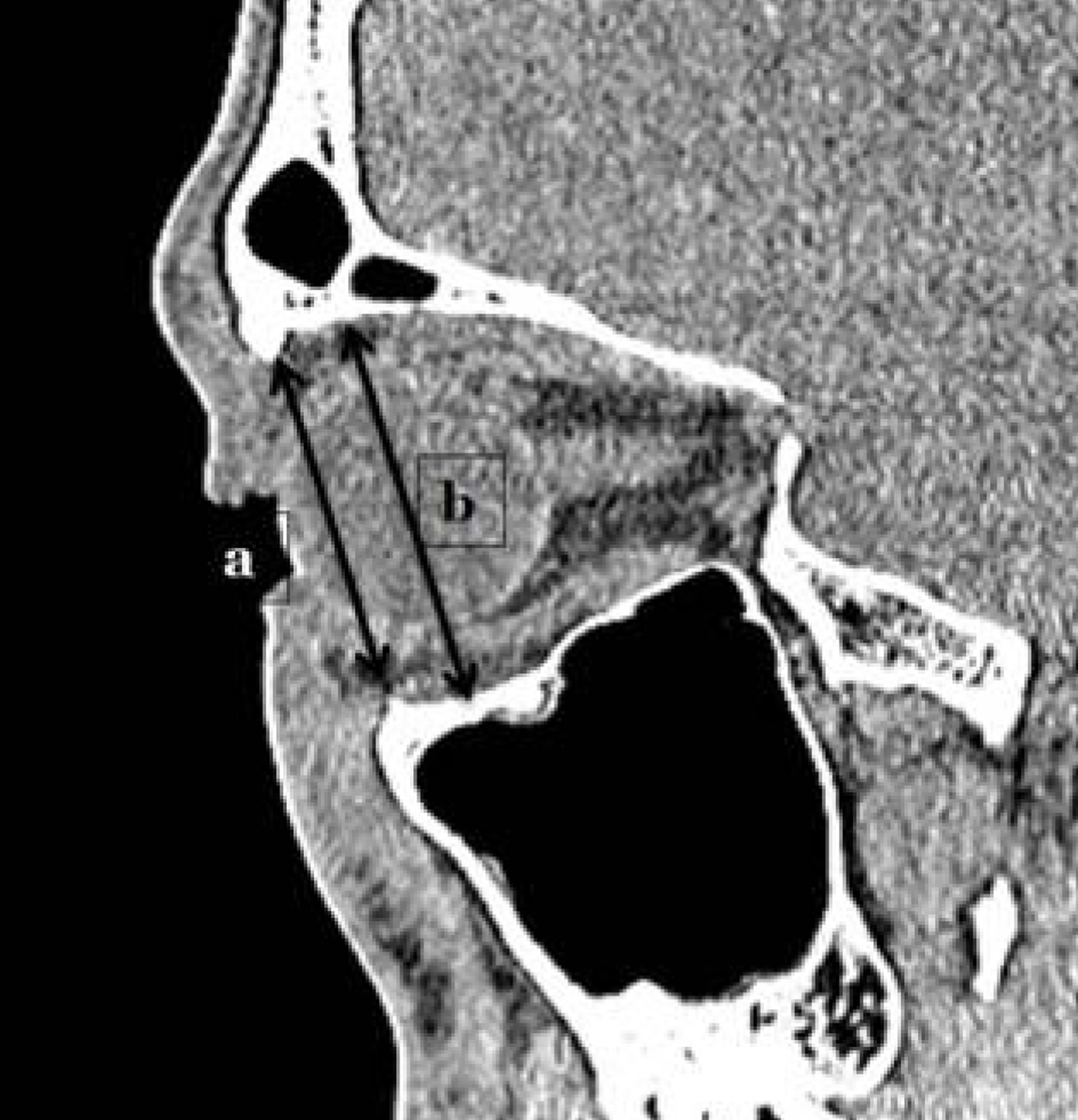
Figure 3. Sagittal computed tomography slice that illustrates 2 length measurements recorded for each orbit. ‘a’ means ante-rior vertical orbital length. ‘b’ means maximal vertical orbital length.
Reference
-
References
1. Hwang K, Baik SH. Surgical anatomy of the orbit of Korean adults. J Craniofac Surg. 1999; 10:129–34.
Article2. Baldeschi L, MacAndie K, Hintschich C. . The removal of the deep lateral wall in orbital decompression: its contribution to exophthalmos reduction and influence on consecutive diplopia. Am J Ophthalmol. 2005; 140:642–7.
Article3. Limawararut V, Valenzuela AA, Sullivan TJ. . Cerebrospinal fluid leaks in orbital and lacrimal surgery. Surv Ophthalmol. 2008; 53:274–84.
Article4. Escaravage GK Jr, Dutton JJ. Age-related changes in the pediatric human orbit on CT. Ophthal Plast Reconstr Surg. 2013; 29:150–6.
Article5. Husmann PR, Samson DR. In the eye of the beholder: sex and race estimation using the human orbital aperture. J Forensic Sci. 2011; 56:1424–9.
Article6. Ozkağ nici A, Büyükmumcu M, Zengin N. . Ocular and peri-orbital anthropometric measurements in term Turkish newborns. Surg Radiol Anat. 2001; 23:321–4.
Article7. Wu KH, Tsai FJ, Li TC. . Normal values of inner canthal dis-tance, interpupillary distance and palpebral fissure length in nor-mal Chinese children in Taiwan. Acta Paediatr Taiwan. 2000; 41:22–7.8. Fok TF, Hon KL, So HK. . Craniofacial anthropometry of Hong Kong Chinese babies: the eye. Orthod Craniofac Res. 2003; 6:48–53.
Article9. Madjarova LM, Madzharov MM, Farkas LG. . Anthropometry of soft-tissue orbits in Bulgarian newborns: norms for intercanthal and biocular widths and length of palpebral fissures in 100 boys and 100 girls. Cleft Palate Craniofac J. 1999; 36:123–6.
Article10. Blake CR, Lai WW, Edward DP. Racial and ethnic differences in-ocular anatomy. Int Ophthalmol Clin. 2003; 43:9–25.11. Leung AK, Ma KC, Siu TO. . Palpebral fissure length. In Chinese newborn infants. Comparison with other ethnic groups. Clin Pediatr (Phila). 1990; 29:172–4.12. Moon D, Wang DH, Park SH. Anthropometric measurements of soft-tissue orbits in Korean newborns. J Korean Ophthalmol Soc. 2012; 53:1385–91.
Article13. Korea Association for Pediatric Ophthalmology and Strabismus. Esodeviation Ⅱ. Current concepts in strabismus. 2nd. Seoul: NaewaeHaksool;2008. p. 194.14. Kang SU, Lee SY, Kim HB, Kim DI. Computed tomographic measurements of the orbit and proptosis in Koreans. J Korean Ophthalmol Soc. 1989; 30:879–86.15. Lim DW, Lee JS, Kim HJ. Normative measurements of Korean or-bital structure. J Korean Ophthalmol Soc. 2001; 42:1–6.
- Full Text Links
-
- Actions
-
Cited
- CITED
-
- Close
- Share
-
- Similar articles
-
- Surgical Anatomy of Deep Lateral Wall by Adults Cadavers and Computed Tomography
- Sex-related and racial variations in orbital floor anatomy
- Orbital Rim Uptake on Bone Scans and Its Clinical Significance
- Analysis of Age-Related Changes in Asian Facial Skeletons Using 3D Vector Mathematics on Picture Archiving and Communication System Computed Tomography
- The Clinical Aspects of Orbital Fractures Proven by Computed Tomography
