J Rhinol.
2017 May;24(1):37-41. 10.18787/jr.2017.24.1.37.
Endoscopic Repair of Spontaneous Cerebrospinal Fluid Rhinorrhea with a Nasoseptal Flap
- Affiliations
-
- 1Department of Otorhinolaryngology-Head and Neck Surgery, Soonchunhyang University College of Medicine, Bucheon, Korea. jyent@schmc.ac.kr
- KMID: 2379788
- DOI: http://doi.org/10.18787/jr.2017.24.1.37
Abstract
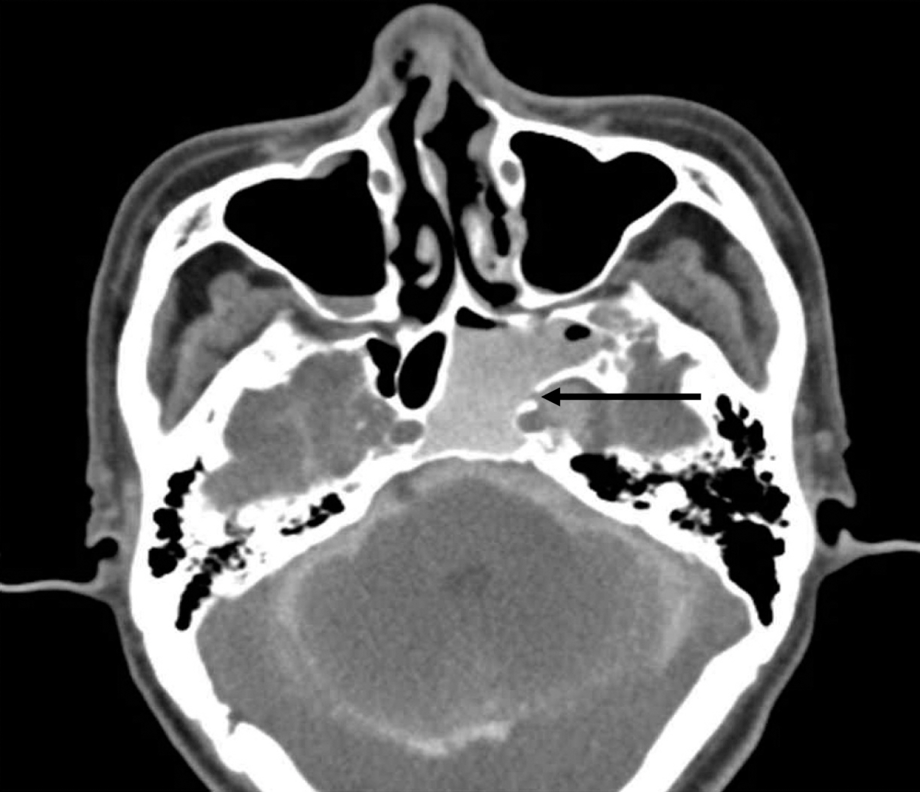
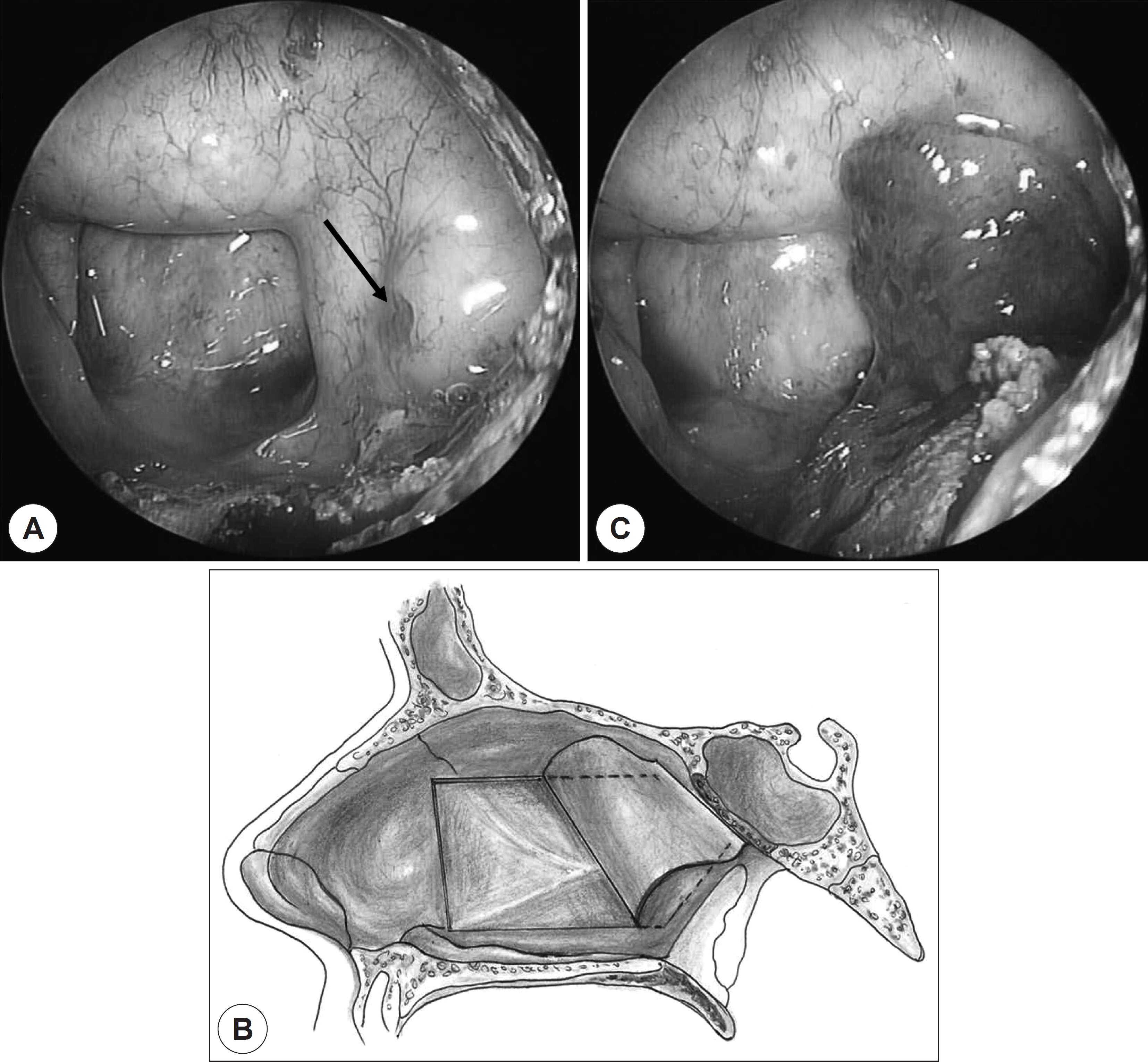
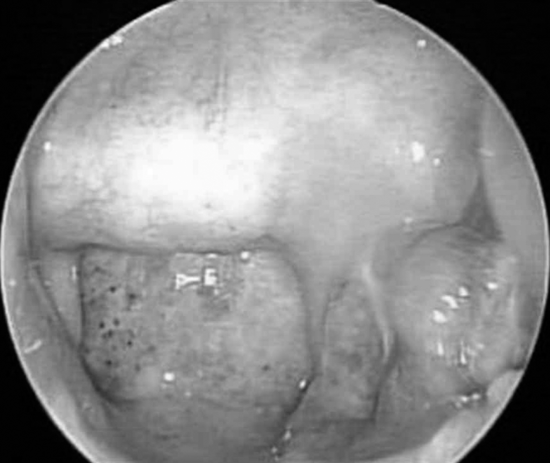
- Cerebrospinal fluid (CSF) rhinorrhea is classified into traumatic and non-traumatic types. Traumatic CSF rhinorrhea comprises the majority of cases, and major causes include head trauma, rhinologic procedures, and neurosurgery. Non-traumatic (spontaneous) CSF rhinorrhea with normal cerebrospinal pressure is a rare condition, occurring in only 4% of cases. We recently experienced a case of spontaneous CSF rhinorrhea complicated with bacterial meningitis. The defect site was identified in the left sphenoid sinus and was successfully repaired with a nasoseptal flap under an endoscopic approach. We present the etiology, classification, and treatment of this rare disease entity with a review of the literature.
Keyword
MeSH Terms
Figure
Reference
-
1). Citardi MJ, Fakhri S. Cerebrospinal fluid rhinorrhea. Flint PW, Haughey BH, Lund V, Niparko JK, Robbins KT, Thomas JR, editors. Cummings Otolaryngology-Head and Neck Surgery. 6th ed.Philadelphia, PA: Elsevier Saunders;2015. p. 803–15.
Article2). Loew F, Pertuiset B, Chaumier EE, Jaksche H. Traumatic spontaneous and postoperative cerebrospinal fluid rhinorrhea. Adv Tech Stand Neurosurg. 1984; 11:169–207.3). Shugar JM, Som PH, Eisman W, Biller HF. Non-traumatic cerebrospinal fluid rhinorrhea. Laryngoscope. 1981; 91(1):114–20.
Article4). Daudia A, Biswas D, Jones NS. Risk of meningitis with cerebrospinal fluid rhinorrhea. Ann Otol Rhinol Laryngol. 2007; 116(12):902–5.
Article5). Katz RT, Kaplan PE. Glucose oxidase sticks and cerebrospinal fluid rhinorrhea. Arch Phys Med Rehabil. 1985; 66(6):391–3.6). berascher G, Arrer E. Efficiency of various methods of identifying cerebrospinal fluid in oto- and rhinorrhea. ORL J Otorhinolaryngol Relat Spec. 1986; 48(6):320–5.7). Lloyd KM, DelGaudio JM, Hudgins PA. Imaging of skull base cerebrospinal fluid leaks in adults. Radiology. 2008; 248(3):725–36.
Article8). Ziu M, Savage JG, Jimenez DF. Diagnosis and treatment of cerebrospinal fluid rhinorrhea following accidental traumatic anterior skull base fractures. Neurosurg Focus. 2012; 32(6):E3.
Article9). Hegazy HM, Carrau RL, Snyderman CH, Kassam A, Zweig J. Trans-nasal endoscopic repair of cerebrospinal fluid rhinorrhea: a meta-analysis. Laryngoscope. 2000; 110(7):1166–72.
Article10). Martin TJ, Loehrl TA. Endoscopic CSF leak repair. Curr Opin Otolaryngol Head Neck Surg. 2007; 15(1):35–9.
Article11). Gassner HG, Ponikau JU, Sherris DA, Kern EB. CSF rhinorrhea: 95 consecutive surgical cases with long term follow-up at the Mayo Clinic. Am J Rhinol. 1999; 13(6):439–47.
Article12). Hadad G, Bassagasteqguy L, Carrau RL, Mataza JC, Kassam AB, Snyderman CH, et al. A novel reconstructive technique after endoscopic expanded endonasal approaches: vascular pedicle nasoseptal flap. Laryngoscope. 2006; 116(10):1882–6.
Article13). Lee KH, Yang CW. Endoscopic endonasal skull base repair with nasoseptal flap. Korean J Otolaryngol. 2015; 58(1):7–11.
Article14). Lopatin AS, Kapitanov DN, Potapov AA. Endonasal endoscopic repair of spontaneous cerebrospinal fluid leaks. Arch Otolaryngol Head Neck Surg. 2003; 129(8):859–63.
Article15). Schlosser RJ, Bolger WE. Nasal cerebrospinal fluid leaks: critical review and surgical considerations. Laryngoscope. 2004; 114(2):255–65.
Article
- Full Text Links
- Actions
-
Cited
- CITED
-
- Close
- Share
- Similar articles
-
- Endoscopic Repair with Above and Below Technique of Recurrent Cerebrospinal Fluid Leak from the Posterior wall of Frontal Sinus: Case Report
- A Case of Encephalomeningocele Presenting with Cerebrospinal Fluid Rhinorrhea: Endoscopic Endonasal Removal with Repair
- Surgical Repair of Cerebrospinal Fluid Rhinorrhea with Mucoperichondrial Free Graft
- Endoscopic Repair of Iatrogenic Cerebrospinal Fluid Rhinorrhea with Osteomucoperiosteal Free Flap Obtained from the Middle Turbinate
- A Case of Endoscopic Repair of Cerebrospinal Fluid Rhinorrhea Associated with Primary Empty Sella Syndrome


