Targeted presurgical decompensation in patients with yaw-dependent facial asymmetry
- Affiliations
-
- 1Department of Orthodontics, Kyung Hee University School of Dentistry, Seoul, Korea. ksj113@khu.ac.kr
- 2Department of Dentistry, Graduate School, Kyung Hee University, Seoul, Korea.
- 3Private Practice, Seoul, Korea.
- 4Department of Orthodontics, School of Dentistry, Seoul National University, Seoul, Korea.
- KMID: 2379653
- DOI: http://doi.org/10.4041/kjod.2017.47.3.195
Abstract
- Facial asymmetry can be classified into the rolling-dominant type (R-type), translation-dominant type (T-type), yawing-dominant type (Y-type), and atypical type (A-type) based on the distorted skeletal components that cause canting, translation, and yawing of the maxilla and/or mandible. Each facial asymmetry type represents dentoalveolar compensations in three dimensions that correspond to the main skeletal discrepancies. To obtain sufficient surgical correction, it is necessary to analyze the main skeletal discrepancies contributing to the facial asymmetry and then the skeletal-dental relationships in the maxilla and mandible separately. Particularly in cases of facial asymmetry accompanied by mandibular yawing, it is not simple to establish pre-surgical goals of tooth movement since chin deviation and posterior gonial prominence can be either aggravated or compromised according to the direction of mandibular yawing. Thus, strategic dentoalveolar decompensations targeting the real basal skeletal discrepancies should be performed during presurgical orthodontic treatment to allow for sufficient skeletal correction with stability. In this report, we document targeted decompensation of two asymmetry patients focusing on more complicated yaw-dependent types than others: Y-type and A-type. This may suggest a clinical guideline on the targeted decompensation in patient with different types of facial asymmetries.
Figure
-
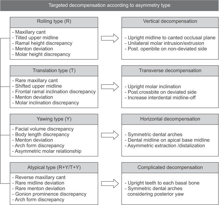
Figure 1 Characteristic and targeted decompensation according to facial asymmetry types.
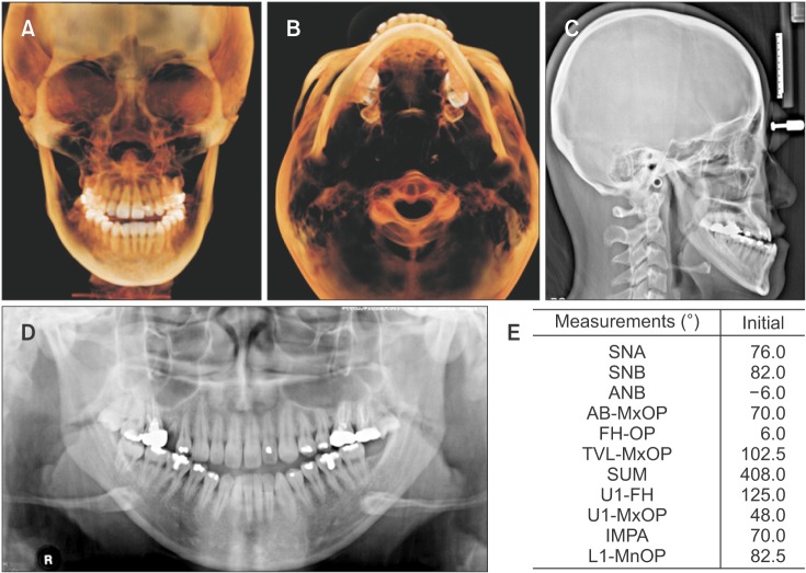
Figure 2 Pretreatment cephalometric and cone-beam computed tomography analyses of a patient (Case 1) with a yawing-dominant type asymmetry. A, Three-dimensional volume-rendering frontal image; B, axial image; C, lateral cephalograph; D, panoramic radiograph; E, cephalometric measurements.Refer to Table 2 for the definitions of each measurement.

Figure 3 Pretreatment photographs of a patient (Case 1) with a yawing-dominant type asymmetry.
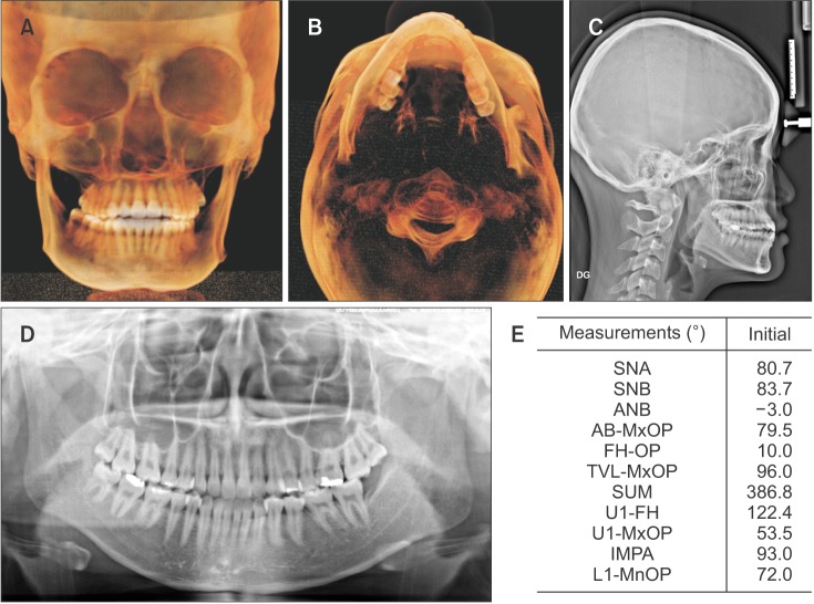
Figure 4 Pretreatment cephalometric and cone-beam computed tomography analyses of a patient (Case 2) with an atypical type asymmetry. A, Three-dimensional volume-rendering frontal image; B, axial image; C, lateral cephalograph; D, panoramic radiograph; E, cephalometric measurements.Refer to Table 2 for the definitions of each measurement.

Figure 5 Pretreatment photographs of a patient (Case 2) with an atypical type asymmetry.
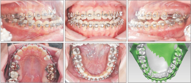
Figure 6 Presurgical intraoral photographs of a patient (Case 1) with a yawing-dominant type asymmetry, and three-dimensional superimposition of the initial and presurgical mandibular models. Gray color, initial mandibular model; green color, pre-surgical mandibular model.
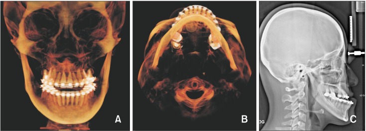
Figure 7 Presurgical three-dimensional volume-rendering frontal (A,) axial images (B,) and lateral cephalograph (C) of a patient (Case 1) with a yawing-dominant type asymmetry.

Figure 8 Presurgical intraoral photographs of a patient (Case 2) with an atypical type asymmetry, and three-dimensional superimposition of the initial and presurgical mandibular models. Gray color, initial mandibular model; green color, presurgical mandibular model.
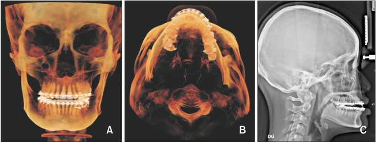
Figure 9 Presurgical three-dimensional volume-rendering frontal (A,) axial images (B,) and lateral cephalograph (C) of a patient (Case 2) with an atypical type asymmetry.
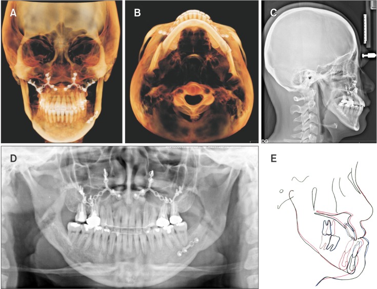
Figure 10 Post-treatmet cephalometric and cone-beam computed tomography images of a patient (Case 1) with a yawing-dominant type asymmetry. A, Three-dimensional volume-rendering frontal image; B, axial image; C, lateral cephalograph; D, panoramic radiograph; E, superim-position (black color, initial; blue color, presurgical; red color, post-treatment).
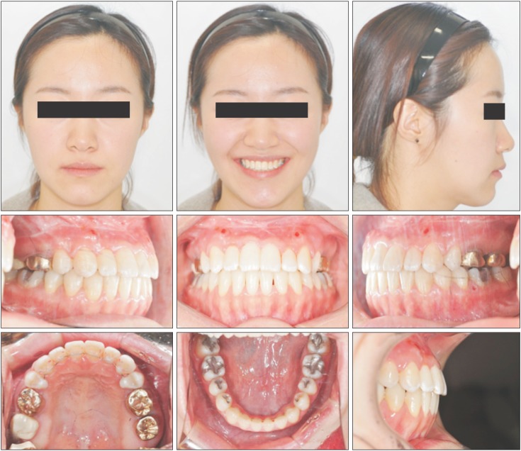
Figure 11 Post-treatment photographs of a patient (Case 1) with a yawing-dominant type asymmetry.
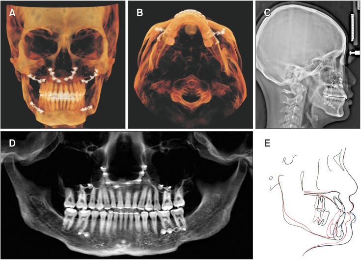
Figure 12 Post-treatmet cephalometric and cone-beam computed tomography images of a patient (Case 2) with an atypical type asymmetry: A, Three-dimensional volume-rendering frontal image; B, axial image; C, lateral cephalograph; D, panoramic radiograph; E, superimposition (black color, initial; blue color, presurgical; red color, post-treatment).
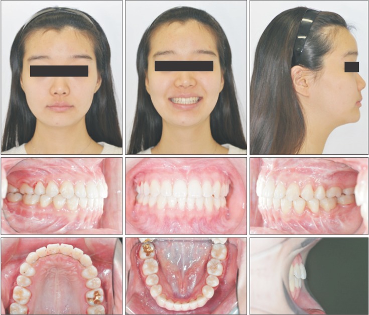
Figure 13 Post-treatment photographs of a patient (Case 2) with an atypical type asymmetry.
Cited by 5 articles
-
Camouflage treatment of posterior bite collapse in a patient with skeletal asymmetry by using posterior maxillary segmental osteotomy
Haitham Badr, Soo-Yeon Lee, Hong-Sik Park, Joo-Young Ohe, Yoon-Goo Kang, Yoon-Goo Kang
Korean J Orthod. 2020;50(4):278-289. doi: 10.4041/kjod.2020.50.4.278.Cone-beam computed tomography analysis of transverse dental compensation in patients with skeletal Class III malocclusion and facial asymmetry
Ji-Yea Lee, Sung-Hoon Han, Hyeong-Seok Ryu, Hee-Min Lee, Sang-Cheol Kim
Korean J Orthod. 2018;48(6):357-366. doi: 10.4041/kjod.2018.48.6.357.Comparison of the three-dimensional structures of mandibular condyles between adults with and without facial asymmetry: A retrospective study
Min-Hee Oh, Sung-Ja Kang, Jin-Hyoung Cho
Korean J Orthod. 2018;48(2):73-80. doi: 10.4041/kjod.2018.48.2.73.Characterization of facial asymmetry phenotypes in adult patients with skeletal Class III malocclusion using three-dimensional computed tomography and cluster analysis
Sang-Woon Ha, Su-Jung Kim, Jin-Young Choi, Seung-Hak Baek
Korean J Orthod. 2022;52(2):85-101. doi: 10.4041/kjod.2022.52.2.85.READER’S FORUM
Hyo-Won Ahn
Korean J Orthod. 2023;53(6):343-344. doi: 10.4041/kjod53.0006RF.
Reference
-
1. Hwang HS, Hwang CH, Lee KH, Kang BC. Maxillofacial 3-dimensional image analysis for the diagnosis of facial asymmetry. Am J Orthod Dentofacial Orthop. 2006; 130:779–785. PMID: 17169741.
Article2. Lee H, Bayome M, Kim SH, Kim KB, Behrents RG, Kook YA. Mandibular dimensions of subjects with asymmetric skeletal class III malocclusion and normal occlusion compared with cone-beam computed tomography. Am J Orthod Dentofacial Orthop. 2012; 142:179–185. PMID: 22858326.
Article3. Baek C, Paeng JY, Lee JS, Hong J. Morphologic evaluation and classification of facial asymmetry using 3-dimensional computed tomography. J Oral Maxillofac Surg. 2012; 70:1161–1169. PMID: 21763045.
Article4. Bruce RA, Hayward JR. Condylar hyperplasia and mandibular asymmetry: a review. J Oral Surg. 1968; 26:281–290. PMID: 4867494.5. Burstone CJ. Diagnosis and treatment planning of patients with asymmetries. Semin Orthod. 1998; 4:153–164. PMID: 9807152.6. Tyan S, Park HS, Janchivdorj M, Han SH, Kim SJ, Ahn HW. Three-dimensional analysis of molar compensation in patients with facial asymmetry and mandibular prognathism. Angle Orthod. 2016; 86:421–430. PMID: 26192894.
Article7. Obwegeser HL, Makek MS. Hemimandibular hyperplasia--hemimandibular elongation. J Maxillofac Surg. 1986; 14:183–208. PMID: 3461097.
Article8. Kusayama M, Motohashi N, Kuroda T. Relationship between transverse dental anomalies and skeletal asymmetry. Am J Orthod Dentofacial Orthop. 2003; 123:329–337. PMID: 12637905.
Article
- Full Text Links
-
- Actions
-
Cited
- CITED
-
- Close
- Share
-
- Similar articles
-
- CAD/CAM splint based on soft tissue 3D simulation for treatment of facial asymmetry
- Characterization of facial asymmetry phenotypes in adult patients with skeletal Class III malocclusion using three-dimensional computed tomography and cluster analysis
- Differences in facial soft tissue deviations in Class III patients with different types of mandibular asymmetry: A cone-beam computed tomography study
- Facial asymmetry
- Evaluation of gonial angle asymmetry after IVRO in facial asymmetry with mandibular prognathism
