A case of prenatally diagnosed extrapulmonary arteriovenous malformation associated with a complex heart defect
- Affiliations
-
- 1Department of Obstetrics and Gynecology, Ajou University School of Medicine, Suwon, Korea.
- 2Department of Obstetrics and Gynecology, University of Ulsan College of Medicine, Asan Medical Center, Seoul, Korea. hswon@amc.seoul.kr
- 3Department of Pathology, University of Ulsan College of Medicine, Asan Medical Center, Seoul, Korea.
- 4Department of Pathology, Samsung Medical Center, Sungkyunkwan University School of Medicine, Seoul, Korea.
- KMID: 2378582
- DOI: http://doi.org/10.5468/ogs.2016.59.6.544
Abstract
- Pulmonary arteriovenous malformations are rare vascular anomalies of the lung, only a few cases of which have been diagnosed prenatally. The diagnostic clue for prenatal diagnosis was cardiomegaly with a particularly enlarged left atrium. All previous cases of pulmonary arteriovenous malformations diagnosed prenatally have been reported as an isolated anomaly or in association with simple heart defects. We here describe the first case of a pulmonary arteriovenous malformation with a complex heart defect that was diagnosed prenatally at 21.0 weeks of gestation and confirmed by postmortem autopsy.
MeSH Terms
Figure
-
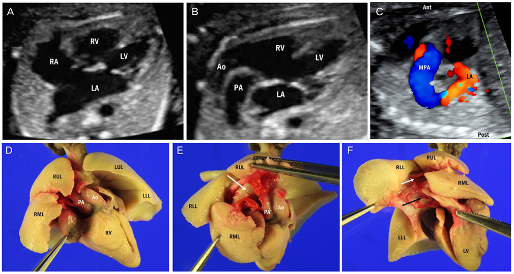
Fig. 1 Fetal echocardiographic images obtained at 21.0 weeks of gestation in the current study case demonstrating cardiomegaly with a particularly enlarged left atrium (LA) in the four-chamber view (A), both aorta (Ao) and pulmonary artery (PA) arising from the right ventricle (RV) (B), and an abnormal communication of main pulmonary artery (MPA) and LA with color Doppler in the parasagittal view (C). Postmortem autopsy showing the abnormal root of Ao in front of the pulmonary artery (PA) arising from the RV in the anterior view (D), PA from the RV running between the right upper lobe (RUL) and the right middle lobe (RML) of the lung turning to the left behind the main bronchus in the right superior view (white arrow, opened PA) (E), and PA connecting to the LA in the right posterior view (white arrow, opened PA; black arrow, opened LA) (F). The inside of LA was dyed by red ink flowing through the PA. RA, right atrium; LV, left ventricle; Ant, anterior; Post, posterior; LUL, left upper lobe; LLL, left lower lobe; RLL, right lower lobe.
Reference
-
1. Akler G, Tamir A, Malinger G, Yagel S. Prenatal diagnosis of a pulmonary arteriovenous malformation. Ultrasound Obstet Gynecol. 2012; 39:235–237.2. Gossage JR, Kanj G. Pulmonary arteriovenous malformations: a state of the art review. Am J Respir Crit Care Med. 1998; 158:643–661.3. Heling KS, Tennstedt C, Goldner B, Bollmann R. Prenatal diagnosis of intrapulmonary arteriovenous malformation: sonographic and pathomorphological findings. Ultrasound Obstet Gynecol. 2002; 19:514–517.4. Kenny DP, Tsai-Goodman B, Martin RP, Tulloh RM. Antenatal diagnosis and postnatal treatment of intrapulmonary arteriovenous malformation. Arch Dis Child Fetal Neonatal Ed. 2007; 92:F366.5. Ostras O, Kurkevych A, Bohuta L, Yalynska T, Raad T, Lewin M, et al. Prenatal diagnosis of left pulmonary artery-to-pulmonary vein fistula and its successful surgical repair in a neonate. Tex Heart Inst J. 2015; 42:169–171.6. Russell MW, Gomez C, Nugent C, Christiansen J. Large fetal pulmonary arteriovenous fistula: impact on pulmonary development. Pediatr Cardiol. 2002; 23:454–457.7. Sinkovskaya E, Berkley E, Bogdan D, Sclater A, Abuhamad A. The role of echocardiography in prenatal diagnosis of pulmonary arteriovenous malformation. Prenat Diagn. 2009; 29:634–636.8. Kalugdan RG, Satoh S, Koyanagi T, Shinzato Y, Nakano H. Antenatal diagnosis of pulmonary arteriovenous fistula using real-time ultrasound and color Doppler flow imaging. J Clin Ultrasound. 1989; 17:607–614.9. Hellmund A, Berg C, Bryan C, Schneider M, Hraska V, Gembruch U. Large fetal pulmonary arteriovenous malformation detected at midtrimester scan with subsequent high cardiac output syndrome and favorable postnatal outcome. Fetal Diagn Ther. 2014; 35:133–136.10. Anabtawi IN, Ellison RG, Ellison LT. Pulmonary arteriovenous aneurysms and fistulas. Anatomical variations, embryology, and classification. Ann Thorac Surg. 1965; 1:277–285.11. Bosher LH Jr, Blake DA, Byrd BR. An analysis of the pathologic anatomy of pulmonary arteriovenous aneurysms with particular reference to the applicability of local excision. Surgery. 1959; 45:91–104.12. Hodgson CH, Kaye RL. Pulmonary arteriovenous fistula and hereditary hemorrhagic telangiectasia: a review and report of 35 cases of fistula. Dis Chest. 1963; 43:449–455.13. Dines DE, Arms RA, Bernatz PE, Gomes MR. Pulmonary arteriovenous fistulas. Mayo Clin Proc. 1974; 49:460–465.14. Gelfand MS, Stephens DS, Howell EI, Alford RH, Kaiser AB. Brain abscess: association with pulmonary arteriovenous fistula and hereditary hemorrhagic Telangiectasia: report of three cases. Am J Med. 1988; 85:718–720.15. Cloutier A, Ash JM, Smallhorn JF, Williams WG, Trusler GA, Rowe RD, et al. Abnormal distribution of pulmonary blood flow after the Glenn shunt or Fontan procedure: risk of development of arteriovenous fistulae. Circulation. 1985; 72:471–479.
- Full Text Links
-
- Actions
-
Cited
- CITED
-
- Close
- Share
-
- Similar articles
-
- Prenatally Diagnosed Extrapulmonary Sequestration: 2cases
- The Management of Arteriovenous Malformation Diagnosed after Extremity Trauma
- A Case of Successful Transarterial Embolization in Arteriovenous Malformation of Uterus
- A Case of Acquired Multiple Arteriovenous Malformations on the Scalp in the Patient of Liver Cirrhosis
- A Giant Aneurysmal Cerebral Arteriovenous Malformation in Childhood: Case Report
