A Case of Coincidental Intrasellar Chordoma and Pituitary Adenoma
- Affiliations
-
- 1Department of Neurosurgery, College of Medicine, Chung-Ang University, Seoul, Korea. kiss798@gmail.com
- 2Department of Pathology, College of Medicine, Chung-Ang University, Seoul, Korea.
- 3Department of Neurosurgery, School of Medicine, Kyungpook National University, Daegu, Korea.
- KMID: 2378307
- DOI: http://doi.org/10.14791/btrt.2017.5.1.49
Abstract
- Although chordomas are midline tumors, primarily intrasellar chordomas are extremely rare. In this report, the authors describe the case of a 68-year-old female with partial abducens nerve palsy in the right eye due to the intrasellar cystic tumor. After endonasal trans-sphenoidal surgery, intraoperative and histopathological findings confirmed the co-occurrence of an entirely intrasellar chordoma and pituitary adenoma. To our knowledge, the present case is the third reported case of an intrasellar chordoma with a pituitary adenoma.
Keyword
MeSH Terms
Figure
-

Fig. 1 Preoperative CT. A: Enhanced brain CT showing a thinned sellar floor and an intrasellar cystic mass compressing the right cavernous sinus without calcification. B: Sagittal images revealing the normal parasellar bony abnormalities.
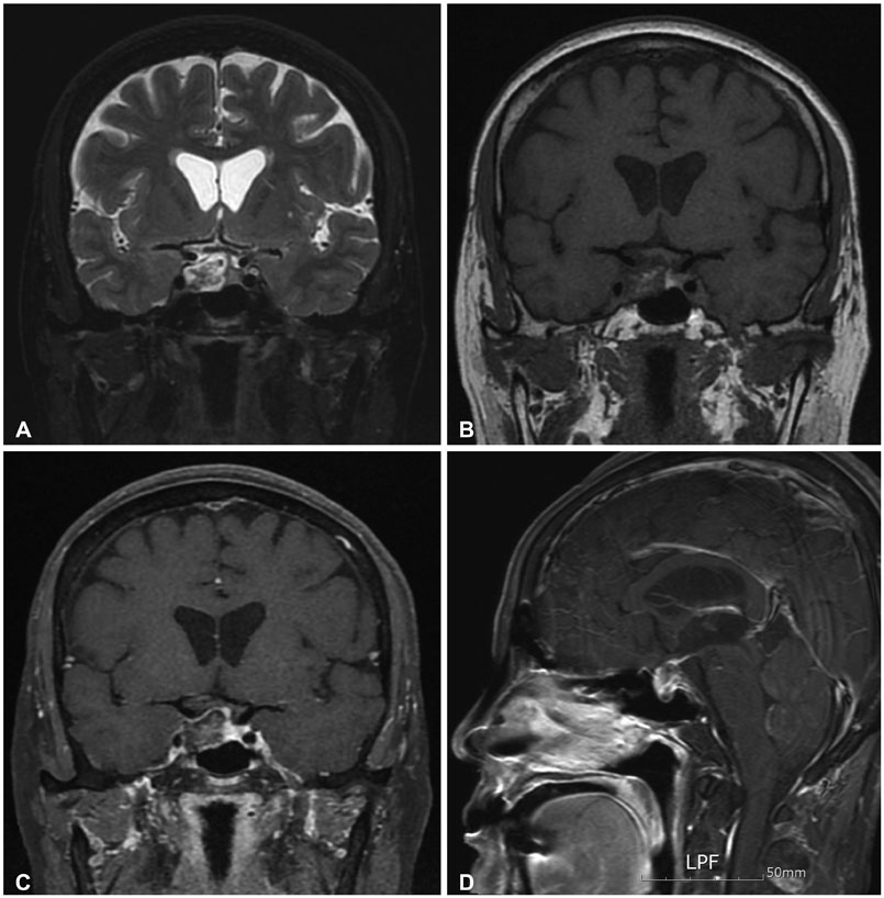
Fig. 2 Preoperative MRI. Brain MRI revealing the intrasellar cystic tumor with a well-enhanced rim and without invasion to the cavernous sinus. A: T2 WI coronal image. B: T1 WI coronal image. C: Enhanced T1 WI coronal image. D: Enhanced T1 WI sagittal image.

Fig. 3 Intraoperative findings showing two different tumors. A: The intact dura and remnant bony sella (arrow). B: A yellowish and friable tumor located to the left and anterior side (arrowheads) and a cystic tumor located to the right side (arrow). C: A prominent yellowish and friable tumor (arrowheads). D: A mucoid substance in the cystic tumor (arrow).
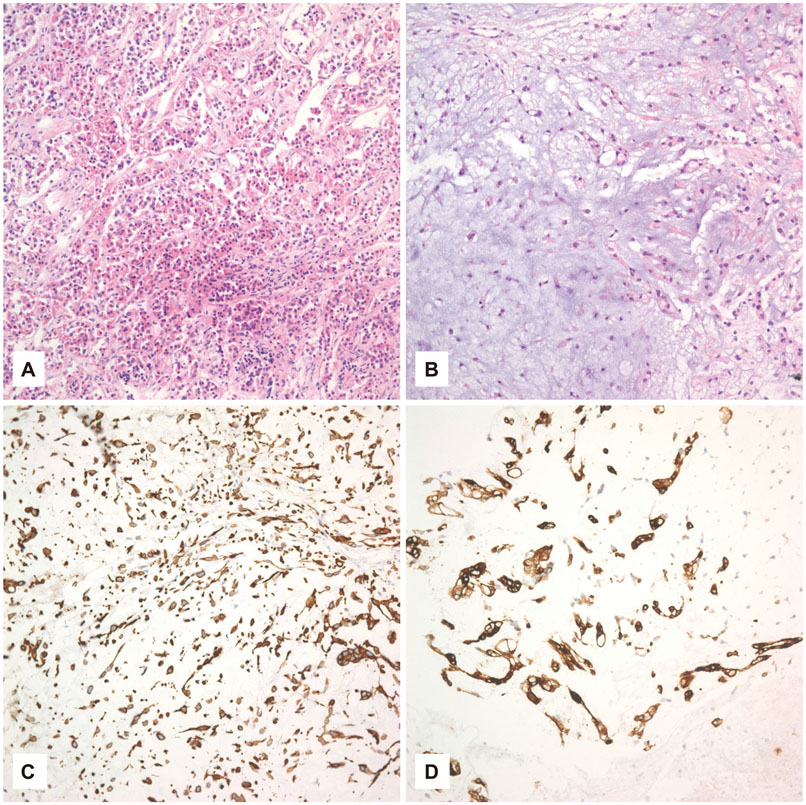
Fig. 4 Histopathological findings of two different tumors. A: Pituitary adenoma: the tumor cells consist of small, oval or polyhedral cells. They are arranged in cords or an acinar pattern. The nuclei of tumor cells are round or oval and contain stippled chromatin (H&E, ×200). B: Chordoma: the neoplasm consists of strands of cells with vacuolated cytoplasm (physaliphorous cells) (H&E, ×200). The neoplastic cells of the chordoma are reactive to the (C) cytokeratin-20 and (D) epithelial membrane antigen antibody in the cytoplasm and cell membrane (×200). H&E, hematoxylin and eosin.
Reference
-
1. Thodou E, Kontogeorgos G, Scheithauer BW, et al. Intrasellar chordomas mimicking pituitary adenoma. J Neurosurg. 2000; 92:976–982.
Article2. Walcott BP, Nahed BV, Mohyeldin A, Coumans JV, Kahle KT, Ferreira MJ. Chordoma: current concepts, management, and future directions. Lancet Oncol. 2012; 13:e69–e76.
Article3. Hirosawa RM, Santos AB, França MM, et al. Intrasellar chondroid chordoma: a case report. ISRN Endocrinol. 2011; 2011:259392.
Article4. Wu AW, Bhuta S, Salamon N, Martin N, Wang MB. Chondroid chordoma of the sella turcica mimicking a pituitary adenoma. Ear Nose Throat J. 2015; 94:E47–E49.5. Kagawa T, Takamura M, Moritake K, Tsutsumi A, Yamasaki T. A case of sellar chordoma mimicking a non-functioning pituitary adenoma with survival of more than 10 years. Noshuyo Byori. 1993; 10:103–106.6. Mathews W, Wilson CB. Ectopic intrasellar chordoma. Case report. J Neurosurg. 1974; 40:260–263.7. Hattori Y, Tahara S, Ishii Y, et al. A case of prolactinoma with chordoma. Clin Neurol Neurosurg. 2013; 115:2537–2539.
Article8. Gökalp HZ, Naderi S. A case of intracranial chordoma associated with pituitary adenoma. J Neurosurg Sci. 1991; 35:103–105.9. Tan WS, Spigos D, Khine N. Chordoma of the sellar region. J Comput Assist Tomogr. 1982; 6:154–158.
Article
