Erdheim-Chester Disease with Emperipolesis: A Unique Case Involving the Heart
- Affiliations
-
- 1Institute of Pathology, Tongji Hospital, Tongji Medical College, Huazhong University of Science and Technology, Wuhan, China. yqduan@hust.edu.cn
- 2Department of Pathology, School of Basic Medical Science, Huazhong University of Science and Technology, Wuhan, China.
- KMID: 2378129
- DOI: http://doi.org/10.4143/crt.2016.078
Abstract
- Histiocytosis is an uncommon disease characterized by excessive accumulation of histiocytes. Here, we report a rare case of non-Langerhans-cell histiocytosis in a 51-year-old woman who presented with severe symptoms of pericardial effusion. Radiologic investigation also detected multiple bone (lower limbs, vertebrae, ribs, and ilium) lesions. Resected pericardium showed abundant mono- or multi-nucleated non-foamy histiocytes (CD68âº/CD163âº/S-100âº/CD1αâ»/langerinâ») in a fibroinflammatory background. The histiocytes demonstrated emperipolesis of lymphocytes, a hallmark feature of Rosai-Dorfman disease (RDD). However, molecular analysis revealed a BRAF V600E mutation of the proliferating histiocytes, highlighting the neoplastic features frequently observed in another non-Langerhans-cell histiocytosis known as Erdheim-Chester Disease (ECD). We consider this case to be a unique presentation of ECD harboring some RDD-like cells with emperipolesis, but not a case of RDD with a BRAF mutation concerning its clinical manifestation (involvement of the heart and bones) and neoplastic features.
MeSH Terms
Figure
-
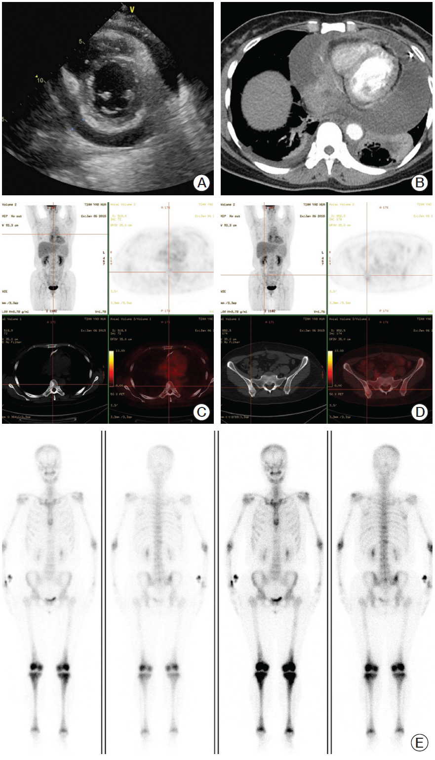
Fig. 1. (A) Ultrasound shows a massive circumferential pericardial effusion. (B) Computerized tomography shows pericardial effusion and pericardial soft tissue density. (C, D) Computerized tomography and positron emission tomography shows radiotracer uptake in the thoracic vertebra (C) and ilium (D). (E) Electrical capacitance tomography demonstrates symmetrical radiotracer uptake in the distal ends of the femurs and the proximal and distal tibia, as well as the ribs and vertebrae.

Fig. 2. (A) The lesion shows infiltration of non-foamy histocytes (arrow) in a marked fibroinflammatory background (H&E staining, ×100). (B) The lesion shows granular histiocytes in a fibroinflammatory background. Emperipolesis (arrow) shows engulfed intact lymphocytes inside the cytoplasm of non-foamy histiocytes (H&E staining, ×200). (C, D) Positive immunostaining of CD68 (C, ×100) and CD163 (D, ×100) in non-foamy histiocytes in a diffuse cytoplasmic pattern. (E) The histiocytes show strong cytoplasmic and nuclear staining for S-100. Engulfed lymphocytes (arrows) are well demonstrated in some histiocytes as emperipolesis (×200). (F) The histiocytes are negative for Langerin (×100).
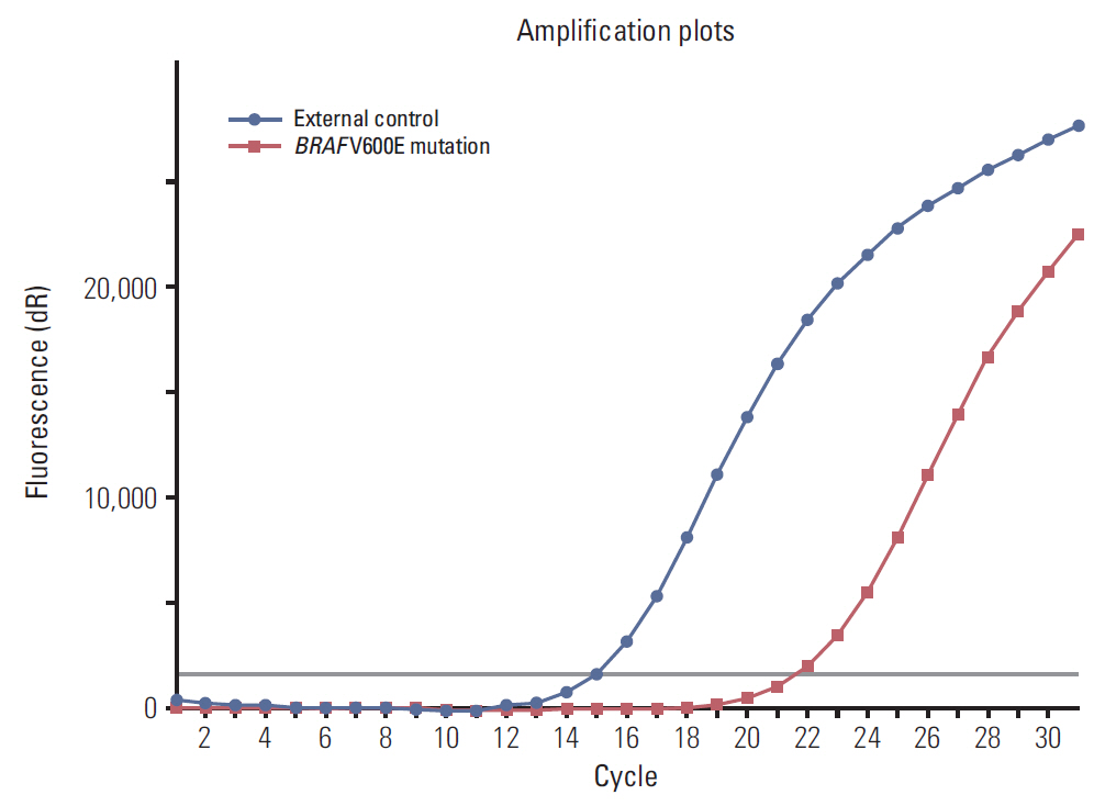
Fig. 3. BRAF–polymerase chain reaction detecting V600E mutation: red amplification curve, BRAF V600E mutation; blue amplification curve, external positive control.
Reference
-
References
1. Emile JF, Abla O, Fraitag S, Horne A, Haroche J, Donadieu J, et al. Revised classification of histiocytoses and neoplasms of the macrophage-dendritic cell lineages. Blood. 2016; 127:2672–81.
Article2. Blombery P, Wong SQ, Lade S, Prince HM. Erdheim-Chester disease harboring the BRAF V600E mutation. J Clin Oncol. 2012; 30:e331.
Article3. Diamond EL, Abdel-Wahab O, Pentsova E, Borsu L, Chiu A, Teruya-Feldstein J, et al. Detection of an NRAS mutation in Erdheim-Chester disease. Blood. 2013; 122:1089–91.
Article4. Diamond EL, Dagna L, Hyman DM, Cavalli G, Janku F, Estrada-Veras J, et al. Consensus guidelines for the diagnosis and clinical management of Erdheim-Chester disease. Blood. 2014; 124:483–92.
Article5. Rastogi V, Sharma R, Misra SR, Yadav L, Sharma V. Emperipolesis: a review. J Clin Diagn Res. 2014; 8:ZM01–2.6. Haroche J, Arnaud L, Amoura Z. Erdheim-Chester disease. Curr Opin Rheumatol. 2012; 24:53–9.
Article7. Satter EK, High WA. Langerhans cell histiocytosis: a review of the current recommendations of the Histiocyte Society. Pediatr Dermatol. 2008; 25:291–5.
Article8. Chen CY, Wu MH, Huang SF, Chen SJ, Lu MY. Langerhans' cell histiocytosis presenting with a para-aortic lesion and heart failure. J Formos Med Assoc. 2001; 100:127–30.9. Foucar E, Rosai J, Dorfman R. Sinus histiocytosis with massive lymphadenopathy (Rosai-Dorfman disease): review of the entity. Semin Diagn Pathol. 1990; 7:19–73.10. Sarraj A, Zarra KV, Jimenez Borreguero LJ, Caballero P, Nuche JM. Isolated cardiac involvement of Rosai-Dorfman disease. Ann Thorac Surg. 2012; 94:2118–20.
Article11. Maleszewski JJ, Hristov AC, Halushka MK, Miller DV. Extranodal Rosai-Dorfman disease involving the heart: report of two cases. Cardiovasc Pathol. 2010; 19:380–4.
Article12. Chen J, Tang H, Li B, Xiu Q. Rosai-Dorfman disease of multiple organs, including the epicardium: an unusual case with poor prognosis. Heart Lung. 2011; 40:168–71.
Article13. Bi Y, Huo Z, Meng Y, Wu H, Yan J, Zhou Y, et al. Extranodal Rosai-Dorfman disease involving the right atrium in a 60-year-old male. Diagn Pathol. 2014; 9:115.
Article14. Lim J, Kim KH, Suh KJ, Yoh KA, Moon JY, Kim JE, et al. A Unique case of Erdheim-Chester disease with axial skeleton, lymph node, and bone marrow involvement. Cancer Res Treat. 2016; 48:415–21.
Article15. Haroche J, Charlotte F, Arnaud L, von Deimling A, Helias-Rodzewicz Z, Hervier B, et al. High prevalence of BRAF V600E mutations in Erdheim-Chester disease but not in other non-Langerhans cell histiocytoses. Blood. 2012; 120:2700–3.
Article
- Full Text Links
-
- Actions
-
Cited
- CITED
-
- Close
- Share
-
- Similar articles
-
- Erdheim-Chester Disease with Perirenal Masses Containing Macroscopic Fat Tissue
- Commentary on "A Case of Erdheim-Chester Disease with Asymptomatic Renal Involvement"
- Erdheim–Chester Disease Involving the Biliary System and Mimicking Immunoglobulin G4-Related Disease: A Case Report
- Reply to Commentary on "A Case of Erdheim-Chester Disease with Asymptomatic Renal Involvement"
- A Case of Erdheim-Chester Disease with Bilateral Hydronephrosis
