Korean Circ J.
2017 Mar;47(2):215-221. 10.4070/kcj.2016.0286.
Safety and Efficacy of an Aortic Arch Stent Graft with Window-Shaped Fenestration for Supra-Aortic Arch Vessels: an Experimental Study in Swine
- Affiliations
-
- 1Division of Cardiology, Department of Internal Medicine, Pusan National University Hospital, Pusan National University College of Medicine, Busan, Korea. glaraone@hanmail.net
- 2Department of Thoracic Surgery, Medical Research Institute, Pusan National University Hospital, Pusan National University College of Medicine, Busan, Korea.
- KMID: 2377462
- DOI: http://doi.org/10.4070/kcj.2016.0286
Abstract
- BACKGROUND AND OBJECTIVES
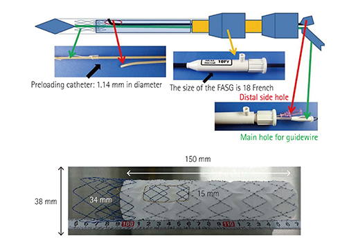
Thoracic endovascular aortic repair exhibits limitations in cases where the aortic pathology involves the aortic arch. We had already developed a fenestrated aortic stent graft (FASG) with a preloaded catheter for aortic pathology involving the aortic arch. FASG was suitable for elective cases.
MATERIALS AND METHODS
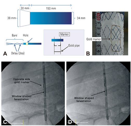
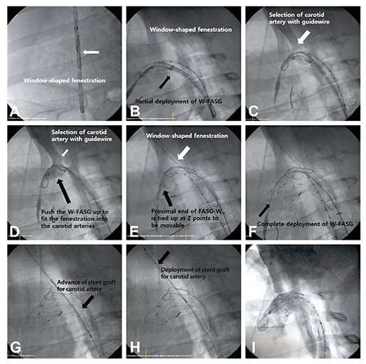
An aortic arch stent graft with a window-shaped fenestration (FASG-W) for supra-aortic arch vessels is suitable for emergent cases. This study aims to test a FASG-W for supra-aortic arch vessels and to perform a preclinical study in swine to evaluate the safety and efficacy of this device. Six FASG-Ws with 1 preloaded catheter were advanced through the iliac artery in 6 swine. The presence of endoleak and the patency and deformity of the grafts were examined with computed tomography (CT) at 4 weeks postoperatively. A postmortem examination was performed at 8 weeks. The mean procedure time for FASG-W was 27.15±4.02 minutes. The mean time for the selection of the right carotid artery was 5.72±0.72 minutes.
RESULTS
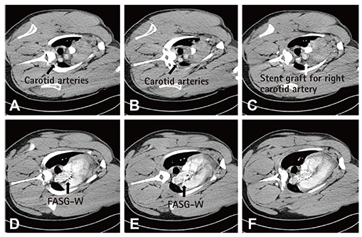
Major adverse events were not observed in any of the 6 pigs who survived for 8 weeks. For the FASG-W, no endoleaks, no disconnection, and no occlusion of the stent grafts were observed in the CT findings or the postmortem gross findings.
CONCLUSION
The procedure with the FASG-W was able to be performed safely in a relatively short procedure time and involved an easy technique. The FASG-W was found to be safe and convenient for use in this preclinical study of swine.
MeSH Terms
Figure
Reference
-
1. Dake MD, Kato N, Mitchell RS, et al. Endovascular stent-graft placement for the treatment of acute aortic dissection. N Engl J Med. 1999; 340:1546–1552.2. Dake MD, Miller DC, Semba CP, Mitchell RS, Walker PJ, Liddell RP. Transluminal placement of endovascular stent-grafts for the treatment of descending thoracic aortic aneurysms. N Engl J Med. 1994; 331:1729–1734.3. Greenberg R, Resch T, Nyman U, et al. Endovascular repair of descending thoracic aortic aneurysms: an early experience with intermediate-term follow-up. J Vasc Surg. 2000; 31(1 Pt 1):147–156.4. Nienaber CA, Rousseau H, Eggebrecht H, et al. Randomized comparison of strategies for type B aortic dissection: the INvestigation of STEnt Grafts in Aortic Dissection (INSTEAD) trial. Circulation. 2009; 120:2519–2528.5. Nienaber CA, Powell JT. Management of acute aortic syndromes. Eur Heart J. 2012; 33:26–35b.6. Shah AA, Barfield ME, Andersen ND, et al. Results of thoracic endovascular aortic repair 6 years after United States Food and Drug Administration approval. Ann Thorac Surg. 2012; 94:1394–1399.7. Zhang H, Wang ZW, Zhou Z, Hu XP, Wu HB, Guo Y. Endovascular stent-graft placement or open surgery for the treatment of acute type B aortic dissection: a meta-analysis. Ann Vasc Surg. 2012; 26:454–461.8. Narayan P, Wong A, Davies I, et al. Thoracic endovascular repair versus open surgical repair - which is the more cost-effective intervention for descending thoracic aortic pathologies? Eur J Cardiothorac Surg. 2011; 40:869–874.9. Makaroun MS, Dillavou ED, Wheatley GH, Cambria RP. Gore TAG Investigators. Five-year results of endovascular treatment with the Gore TAG device compared with open repair of thoracic aortic aneurysms. J Vasc Surg. 2008; 47:912–918.10. Lee SH, Chung CH, Jung SH, et al. Midterm outcomes of open surgical repair compared with thoracic endovascular repair for isolated descending thoracic aortic disease. Korean J Radiol. 2012; 13:476–482.11. Al-Nouri O, Moeller C, Borrowdale R, Milner R. Late complication of thoracic endovascular stent-grafting. Ann Vasc Surg. 2011; 25:982.e1–982.e4.12. Lee CJ, Rodriguez HE, Kibbe MR, Malaisrie SC, Eskandari MK. Secondary interventions after elective thoracic endovascular aortic repair for degenerative aneurysms. J Vasc Surg. 2013; 57:1269–1274.13. Ullery BW, McGarvey M, Cheung AT, et al. Vascular distribution of stroke and its relationship to perioperative mortality and neurologic outcome after thoracic endovascular aortic repair. J Vasc Surg. 2012; 56:1510–1517.14. Grabenwöger M, Alfonso F, Bachet J, et al. European Association for Cardio-Thoracic Surgery (EACTS). European Society of Cardiology (ESC). European Association of Percutaneous Cardiovascular Interventions (EAPCI). Thoracic Endovascular Aortic Repair (TEVAR) for the treatment of aortic diseases: a position statement from the European Association for Cardio-Thoracic Surgery (EACTS) and the European Society of Cardiology (ESC), in collaboration with the European Association of Percutaneous Cardiovascular Interventions (EAPCI). Eur J Cardiothorac Surg. 2012; 42:17–24.15. Lee KN, Lee HC, Park JS, et al. The modified chimney technique with a thoracic aortic stent graft to preserve the blood flow of the left common carotid artery for treating descending thoracic aortic aneurysm and dissection. Korean Circ J. 2012; 42:360–365.16. Sun Z, Mwipatayi BP, Allen YB, Hartley DE, Lawrence-Brown MM. Multislice CT angiography of fenestrated endovascular stent grafting for treating abdominal aortic aneurysms: a pictorial review of the 2D/3D visualizations. Korean J Radiol. 2009; 10:285–293.17. Inoue K, Hosokawa H, Iwase H, et al. Aortic arch reconstruction by transluminally placed endovascular branched stent graft. Circulation. 1999; 100:19 Suppl. II316–II321.18. Schneider DB, Curry TK, Reilly LM, Kang JW, Messina LM, Chuter TA. Branched endovascular repair of aortic arch aneurysm with a modular stent-graft system. J Vasc Surg. 2003; 38:855.19. Ahanchi SS, Almaroof B, Stout CL, Panneton JM. In situ laser fenestration for revascularization of the left subclavian artery during emergent thoracic endovascular aortic repair. J Endovasc Ther. 2012; 19:226–230.20. Wei G, Xin J, Yang D, et al. A new modular stent graft to reconstruct aortic arch. Eur J Vasc Endovasc Surg. 2009; 37:560–565.21. Kim SP, Lee HC, Park TS, et al. Safety and efficacy of a novel, fenestrated aortic arch stent graft with a preloaded catheter for supraaortic arch vessels: an experimental study in Swine. J Korean Med Sci. 2015; 30:426–434.
- Full Text Links
- Actions
-
Cited
- CITED
-
- Close
- Share
- Similar articles
-
- Hybrid Procedure for Aortic Arch Repair: Arch Vessels Debranching with Supraaortic Revascularization Followed by Endovascular Aortic Stent Grafting
- Hybrid Method for Stent-graft Insertion in a Patient with a Thoracic Aortic Aneurysm Involving the Aortic Arch: A case report
- Safety and Efficacy of a Novel, Fenestrated Aortic Arch Stent Graft with a Preloaded Catheter for Supraaortic Arch Vessels: An Experimental Study in Swine
- Successful Treatment of a Ruptured Aortic Arch Aneurysm Using a Hybrid Procedure
- Aortic Arch Debranching and Antegrade Stent Graft Placement in an Expanding Distal Dissecting Aneurysm after Repair of an Acute Type I Aortic Dissection


