Full mouth rehabilitation of edentulous patient with intellectual disability using implants and monolithic zirconia
- Affiliations
-
- 1Department of Prosthodontics and Dental Research Institute, School of Dentistry, Seoul National University, Seoul, Republic of Korea. ksh1250@snu.ac.kr
- KMID: 2377184
- DOI: http://doi.org/10.4047/jkap.2017.55.2.156
Abstract
- People with class I intellectual disability need lifelong assistance and protection from their surroundings due to impaired adaptive functioning. They have poor oral health and show higher prevalence of dental caries, periapical inflammation and tooth loss that require proper prosthetic restoration. Because removable prostheses for intellectually disabled patients often lack stability, retention, and maintenance, fixed prostheses are essential and the only available option is dental implants. In this case, a 45 year-old male patient with class I intellectual disability had poor oral hygiene with most of his teeth missing and visited the clinic to recover his masticatory function. Due to such systemic conditions, the definitive restoration of choice was the implant-supported fixed dental prosthesis made of biocompatible and highly strong monolithic zirconia. In consequence of the treatment process, the patient was able to improve his oral environment aesthetically and functionally.
MeSH Terms
Figure
-
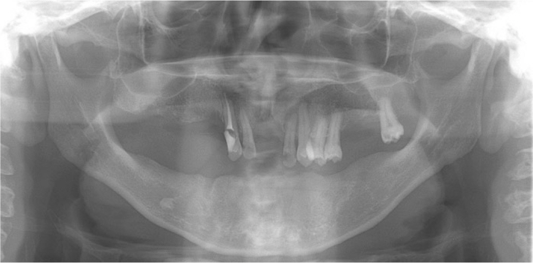
Fig. 1. Initial intraoral photographs. (A) Maxillary occlusal view, (B) Frontal view, (C) Mandibular occlusal view.
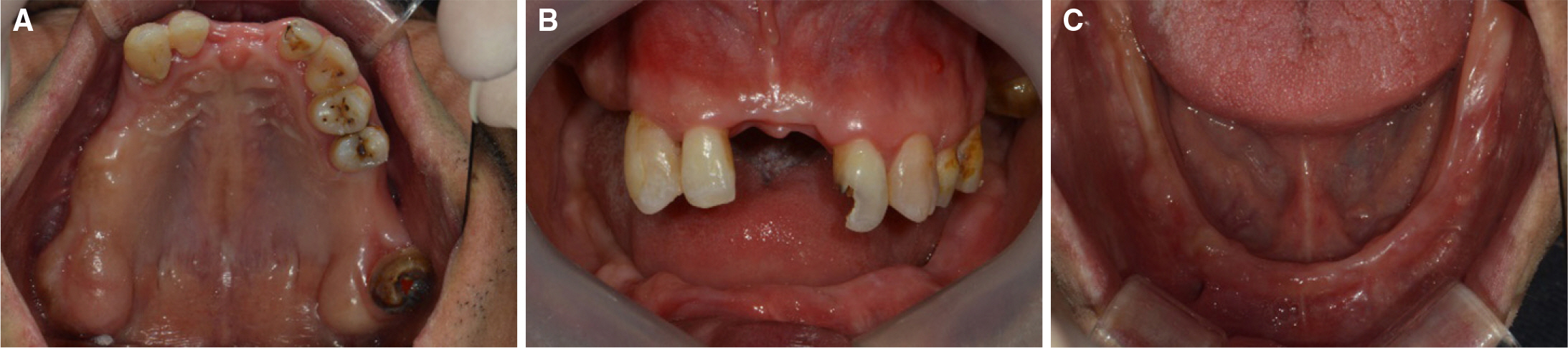
Fig. 2. Initial panoramic radiograph of the patient.
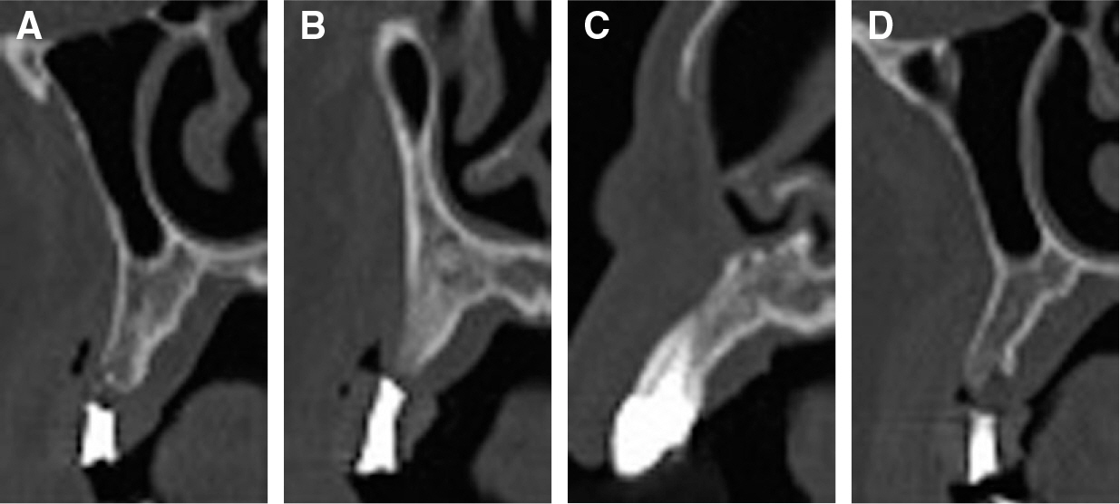
Fig. 3. The CBCT images at the maxilla. (A) #16, (B) #15, (C) #13, (D) #26.
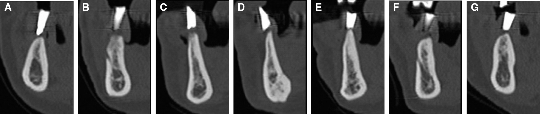
Fig. 4. The CBCT images at the mandible. (A) #46, (B) #44, (C) #43, (D) #31, (E) #33, (F) #34, (G) #36.
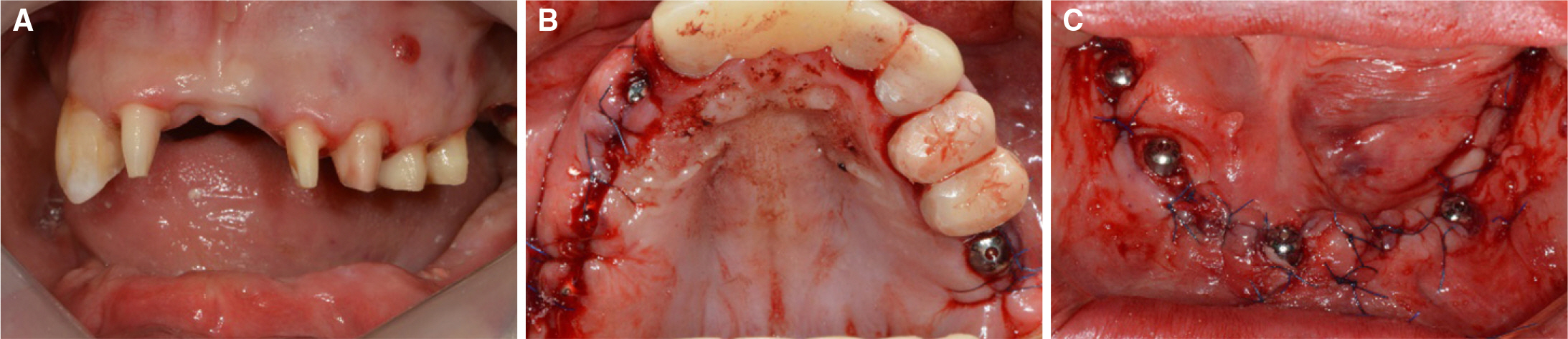
Fig. 5. The surgical manipulations performed under general anesthesia. (A) Tooth preparation, (B) Implant surgery (Maxilla), (C) Implant surgery (Mandible).
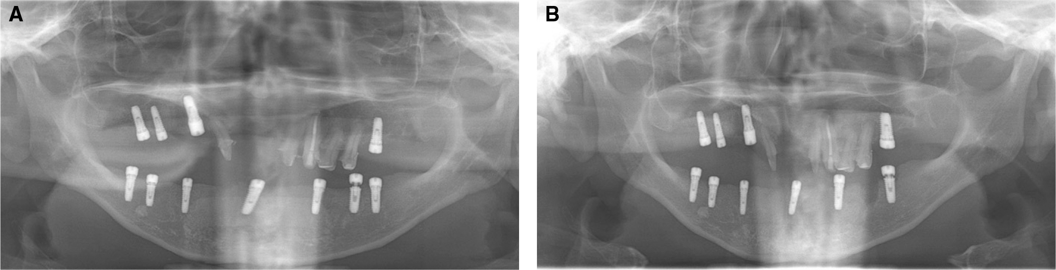
Fig. 6. Radiographs of the patient. (A) Immediately after surgery, (B) After 1 month.
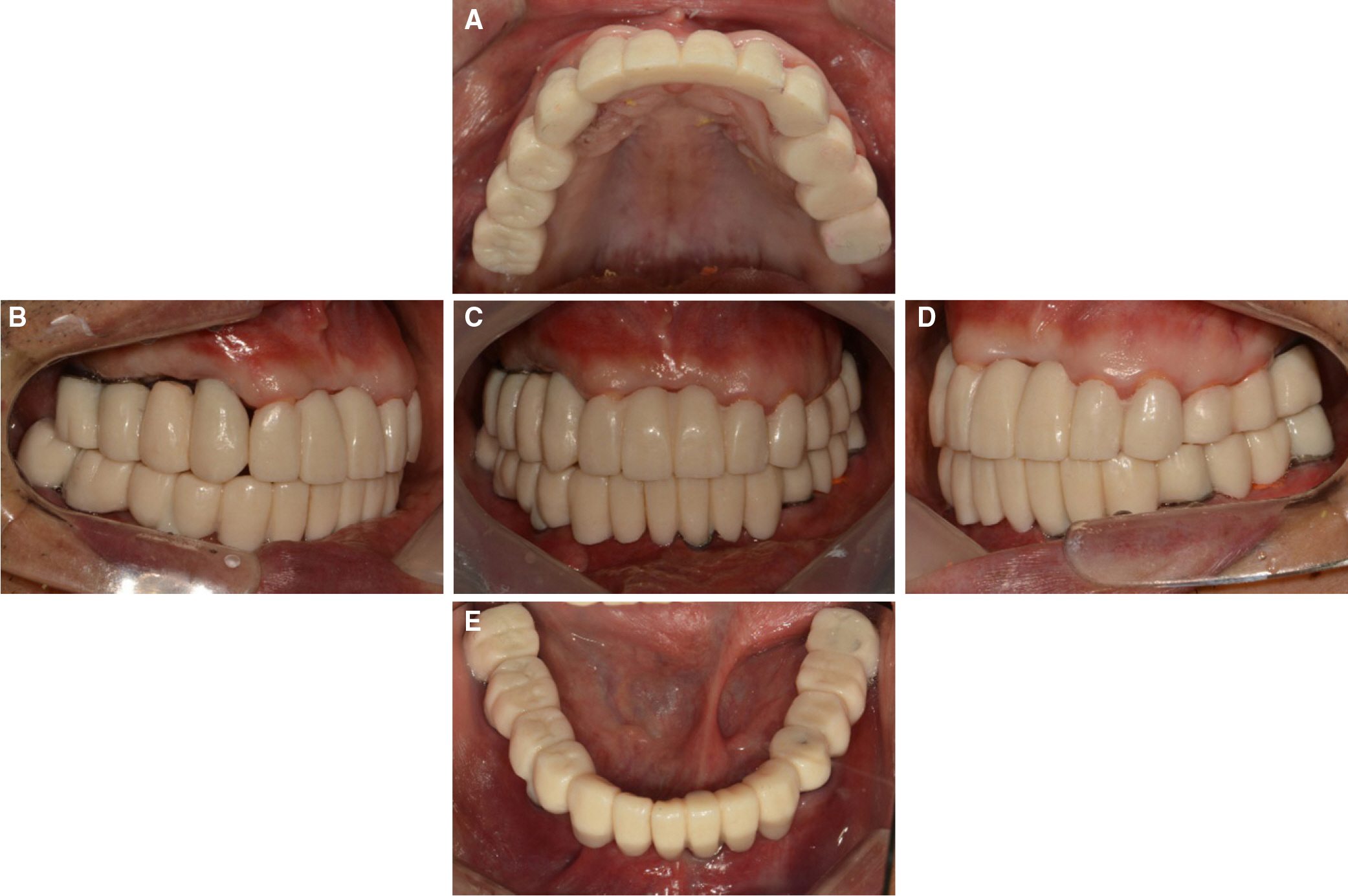
Fig. 7. Provisional prostheses. (A) Maxillary occlusal view, (B) Lateral view (right), (C) Frontal view, (D) Lateral view (left), (E) Mandibular occlusal view.
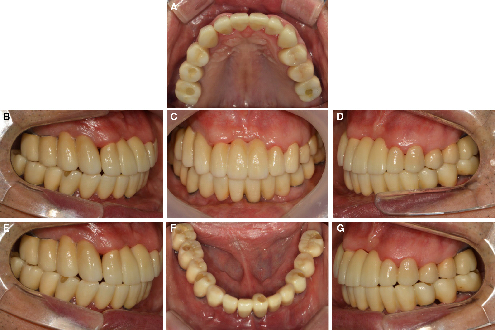
Fig. 8. Definitive prostheses (CAD/CAM monolithic zirconia). (A) Maxillary occlusal view, (B) Lateral view (right), (C) Frontal view, (D) Lateral view (left), (E) Right working movement, (F) Mandibular occlusal view, (G) Left working movement.
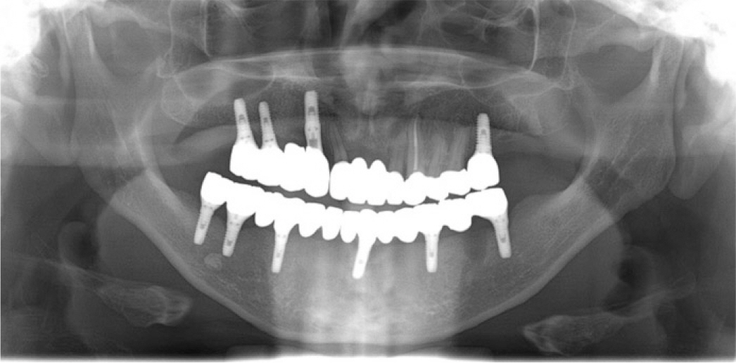
Fig. 9. Panoramic radiograph of the patient after treatment.
Reference
-
1.Moon SY., Kim SK. Implants in psychiatric patients. J Korean Dis Oral Health. 2007. 3:1–5.2.Scully C., Hobkirk J., Dios PD. Dental endosseous implants in the medically compromised patient. J Oral Rehabil. 2007. 34:590–9.
Article3.Diz P., Scully C., Sanz M. Dental implants in the medically compromised patient. J Dent. 2013. 41:195–206.
Article4.Isaksson R., Becktor JP., Brown A., Laurizohn C., Isaksson S. Oral health and oral implant status in edentulous patients with implant-supported dental prostheses who are receiving long-term nursing care. Gerodontology. 2009. 26:245–9.
Article5.Lustig JP., Yanko R., Zilberman U. Use of dental implants in patients with Down syndrome: a case report. Spec Care Dentist. 2002. 22:201–4.
Article6.Cune MS., Strooker H., van der Reijden WA., de Putter C., Laine ML., Verhoeven JW. Dental implants in persons with severe epilepsy and multiple disabilities: a long-term retrospective study. Int J Oral Maxillofac Implants. 2009. 24:534–40.7.Ribeiro CG., Siqueira AF., Bez L., Cardoso AC., Ferreira CF. Dental implant rehabilitation of a patient with down syndrome: a case report. J Oral Implantol. 2011. 37:481–7.
Article8.Feijoo JF., Limeres J., Diniz M., Del Llano A., Seoane J., Diz P. Osseointegrated dental implants in patients with intellectual disability: a pilot study. Disabil Rehabil. 2012. 34:2025–30.
Article9.Lee BC., Jung GY., Kim DJ., Han JS. Initial bacterial adhesion on resin, titanium and zirconia in vitro. J Adv Prosthodont. 2011. 3:81–4.10.Triwatana P., Nagaviroj N., Tulapornchai C. Clinical performance and failures of zirconia-based fixed partial dentures: a review literature. J Adv Prosthodont. 2012. 4:76–83.
Article11.You JW., Heo SJ., Kim SK., Koak JY. Full mouth rehabilitation of an oligodontia patient with intellectual disability based on shortened dental arch concept: a case report. J Korean Acad Prosthodont. 2012. 50:330–5.
Article12.Solow RA. Comprehensive implant restoration and the shortened dental arch. Gen Dent. 2010. 58:390–9.13.Kim HY., Yeo IS., Lee JB., Kim SH., Kim DJ., Han JS. Initial in vitro bacterial adhesion on dental restorative materials. Int J Artif Organs. 2012. 35:773–79.
Article14.Fischman B. The rotational aspect of mandibular flexure. J Prosthet Dent. 1990. 64:483–5.
Article15.Canabarro Sde A., Shinkai RS. Medial mandibular flexure and maximum occlusal force in dentate adults. Int J Prosthodont. 2006. 19:177–82.16.Zarone F., Apicella A., Nicolais L., Aversa R., Sorrentino R. Mandibular flexure and stress build-up in mandibular full-arch fixed prostheses supported by osseointegrated implants. Clin Oral Implants Res. 2003. 14:103–14.
Article17.Paez CY., Barco T., Roushdy S., Andres C. Split-frame implant prosthesis designed to compensate for mandibular flexure: a clinical report. J Prosthet Dent. 2003. 89:341–3.
Article18.Schmitter M., Mussotter K., Rammelsberg P., Stober T., Ohlmann B., Gabbert O. Clinical performance of extended zirconia frameworks for fixed dental prostheses: two-year results. J Oral Rehabil. 2009. 36:610–5.
Article19.Tinschert J., Schulze KA., Natt G., Latzke P., Heussen N., Spiekermann H. Clinical behavior of zirconia-based fixed partial dentures made of DC-Zirkon: 3-year results. Int J Prosthodont. 2008. 21:217–22.
- Full Text Links
-
- Actions
-
Cited
- CITED
-
- Close
- Share
-
- Similar articles
-
- Full mouth rehabilitation using monolithic zirconia: a clinical report
- Full mouth rehabilitation of a patient using monolithic zirconia and dental CAD/CAM system: a case report
- Full-mouth rehabilitation with implant-supported fixed dental prostheses for the edentulous maxilla and partially edentulous mandible: A case report
- A case of full mouth rehabilitation with orthodontic treatment in patient with extensive tooth erosion and wear using monolithic zirconia prostheses
- Maxillary cement retained implant supported monolithic zirconia prosthesis in a full mouth rehabilitation: a clinical report
