J Adv Prosthodont.
2016 Jun;8(3):201-206. 10.4047/jap.2016.8.3.201.
Comparative study of the radiopacity of resin cements used in aesthetic dentistry
- Affiliations
-
- 1Department of Restorative Dentistry, Faculty of Dentistry, Catholic University of Valencia, Valencia, Spain. raquelmontesfariza@gmail.com
- KMID: 2376844
- DOI: http://doi.org/10.4047/jap.2016.8.3.201
Abstract
- PURPOSE
The aim of this study was to compare the radiopacity of 6 modern resin cements with that of human enamel and dentine using the Digora digital radiography system, to verify whether they meet the requirements of ANSI/ADA specification no. 27/1993 and the ISO 4049/2000 standard and assess whether their radiopacity is influenced by the thickness of the cement employed.
MATERIALS AND METHODS
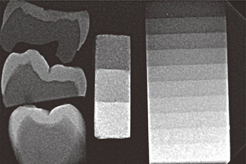
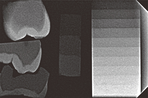
Three 3-thickness samples (0.5, 1 and 1.5 mm) were fabricated for each material. The individual cement samples were radiographed on the CCD sensor next to the aluminium wedge and the tooth samples. Five radiographs were made of each sample and therefore five readings of radiographic density were taken for each thickness of the materials. The radiopacity was measured in pixels using Digora 2.6 software. The calibration curve obtained from the mean values of each step of the wedge made it possible to obtain the equivalent in mm of aluminium for each mm of the luting material.
RESULTS
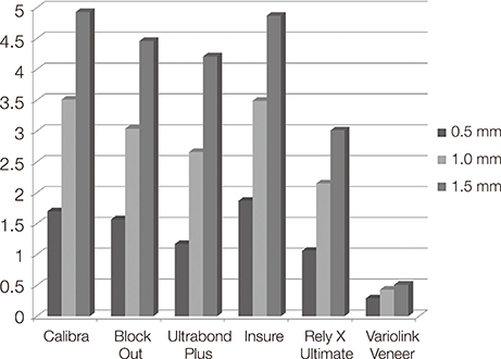
With the exception of Variolink Veneer Medium Value 0, all the cements studied were more radiopaque than enamel and dentin (P<.05) and complied with the ISO and ANSI/ADA requirements (P<.001). The radiopacity of all the cements examined depended on their thickness: the thicker the material, the greater its radiopacity.
CONCLUSION
All materials except Variolink Veneer Medium Value 0 yielded radiopacity values that complied with the recommendations of the ISO and ANSI/ADA. Variolink Veneer Medium Value 0 showed less radiopacity than enamel and dentin.
Keyword
MeSH Terms
Figure
Reference
-
1. Ladha K, Verma M. Conventional and contemporary luting cements: an overview. J Indian Prosthodont Soc. 2010; 10:79–88.2. Fernandes LO, Cassimiro-Silva PF, Massa ME, Pedrosa RF, Cardoso SMO. Radiopacity of resin cements: in vitro study. Rev Gaúcha Odontol. 2012; 60:169–177.3. Reis JM, Jorge EG, Ribeiro JG, Pinelli LA, Abi-Rached Fde O, Tanomaru-Filho M. Radiopacity evaluation of contemporary luting cements by digitization of images. ISRN Dent. 2012; 2012:704246.4. Tsuge T. Radiopacity of conventional, resin-modified glass ionomer, and resin-based luting materials. J Oral Sci. 2009; 51:223–230.5. Rosenstiel SF, Land MF, Crispin BJ. Dental luting agents: A review of the current literature. J Prosthet Dent. 1998; 80:280–301.6. Altintas SH, Yildirim T, Kayipmaz S, Usumez A. Evaluation of the radiopacity of luting cements by digital radiography. J Prosthodont. 2013; 22:282–286.7. Dukic W, Delija B, Derossi D, Dadic I. Radiopacity of composite dental materials using a digital X-ray system. Dent Mater J. 2012; 31:47–53.8. Gürdal P, Akdeniz BG. Comparison of two methods for radiometric evaluation of resin-based restorative materials. Dentomaxillofac Radiol. 1998; 27:236–239.9. Pekkan G, Ozcan M. Radiopacity of different resin-based and conventional luting cements compared to human and bovine teeth. Dent Mater J. 2012; 31:68–75.10. Pedrosa RF, Brasileiro IV, dos Anjos Pontual ML, dos Anjos Pontual A, da Silveira MM. Influence of materials radiopacity in the radiographic diagnosis of secondary caries: evaluation in film and two digital systems. Dentomaxillofac Radiol. 2011; 40:344–350.11. Salzedas LM, Louzada MJ, de Oliveira Filho AB. Radiopacity of restorative materials using digital images. J Appl Oral Sci. 2006; 14:147–152.12. Fonseca RB, Branco CA, Soares PV, Correr-Sobrinho L, Haiter-Neto F, Fernandes-Neto AJ, Soares CJ. Radiodensity of base, liner and luting dental materials. Clin Oral Investig. 2006; 10:114–118.13. Fraga RC, Luca-Fraga LR, Pimenta LA. Physical properties of resinous cements: an in vitro study. J Oral Rehabil. 2000; 27:1064–1067.14. Hill EE. Dental cements for definitive luting: a review and practical clinical considerations. Dent Clin North Am. 2007; 51:643–658.15. Pegoraro TA, da Silva NR, Carvalho RM. Cements for use in esthetic dentistry. Dent Clin North Am. 2007; 51:453–471.16. Attar N, Tam LE, McComb D. Mechanical and physical properties of contemporary dental luting agents. J Prosthet Dent. 2003; 89:127–134.17. Manso AP, Silva NR, Bonfante EA, Pegoraro TA, Dias RA, Carvalho RM. Cements and adhesives for all-ceramic restorations. Dent Clin North Am. 2011; 55:311–332.18. el-Mowafy O. The use of resin cements in restorative dentistry to overcome retention problems. J Can Dent Assoc. 2001; 67:97–102.19. Van Dijken JW, Wing KE, Ruyter IE. An evaluation of the radiopacity of composite restorative materials used in class I and class II cavities. Acta Odontol Scand. 1989; 47:401–407.20. Furtos G, Baldea B, Silaghi-Dumitrescu L, Moldovan M, Prejmerean C, Nica L. Influence of inorganic filler content on the radiopacity of dental resin cements. Dent Mater J. 2012; 31:266–272.21. American National Standard/American Dental Association. Proposed ANSI/ADA Specification 27, Dentistry - Polymer-based filling. Restorative and Luting Materials, July 2005. Chicago: ADA.22. ISO standard 4049. Dentistry-polymer-based filling, restorative and luting materials. Geneva; Switzerland: International Organization for Standardization;2000. p. 1–27.23. Gu S, Rasimick BJ, Deutsch AS, Musikant BL. Radiopacity of dental materials using a digital X-ray system. Dent Mater. 2006; 22:765–770.
- Full Text Links
- Actions
-
Cited
- CITED
-
- Close
- Share
- Similar articles
-
- Simple methods to enhance bond strength of self-adhesive resin cements
- Light curing of dual cure resin cement
- 'Wet or Dry tooth surface?' - for self-adhesive resin cement
- The film thickness and retention of cast crown using adhesive resin cements
- A study on the bond strength of porcelain laminate and composite resin cements


