J Adv Prosthodont.
2017 Apr;9(2):93-98. 10.4047/jap.2017.9.2.93.
Analysis of the width ratio and wear rate of maxillary anterior teeth in the Korean population
- Affiliations
-
- 1Department of Prosthodontics, School of Dentistry, Chonnam National University, Gwangju, Republic of Korea. upgradepc@hanmail.net
- KMID: 2376295
- DOI: http://doi.org/10.4047/jap.2017.9.2.93
Abstract
- PURPOSE
The purpose of this study was to compare the width ratio of maxillary anterior teeth according to age in the Korean population and to evaluate the maxillary central incisor width-to-length (W/L) ratio, given differences in age and gender.
MATERIALS AND METHODS
Ninety-three Korean adults were divided into 3 groups (n = 31) by age. Group I was 20 - 39 years old, Group II was 40 - 59 years old, and Group III was over 60 years of age. After taking an impression and a cast model of the maxillary arch, the anterior teeth width ratio and central incisor W/L ratio were calculated from standard digital images of the cast models using a graph paper with a digital single lens reflex (DSLR) camera. The calculated ratios were compared among all groups and central incisor W/L ratio were analyzed according to age and gender. All comparative data were statistically analyzed with one-sample t-tests, one-way ANOVAs with Tukey tests, and independent t-tests.
RESULTS
No significant differences in maxillary anterior teeth ratios were found among the age groups. The maxillary central incisor W/L ratios in Group III were the greatest and were significantly higher than those in the other groups. The central incisor W/L ratio of men was higher than that of women in Group II.
CONCLUSION
Maxillary anterior teeth width ratios were similar in all age groups in the Korean population. The maxillary central incisor was observed as worn teeth in the group over 60 years of age, and a significant difference between genders was found in 40 to 50 year olds.
Keyword
Figure
-
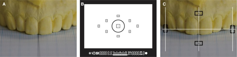
Fig. 1 (A) Maxillary dental cast setting on graph paper, (B) DSLR camera's viewfinder, (C) Photographing view by auto focusing mode with 4 points.

Fig. 2 The visible mesio-distal width of maxillary anterior teeth was measured at the central incisor (A), lateral incisor (B), and canine (C).
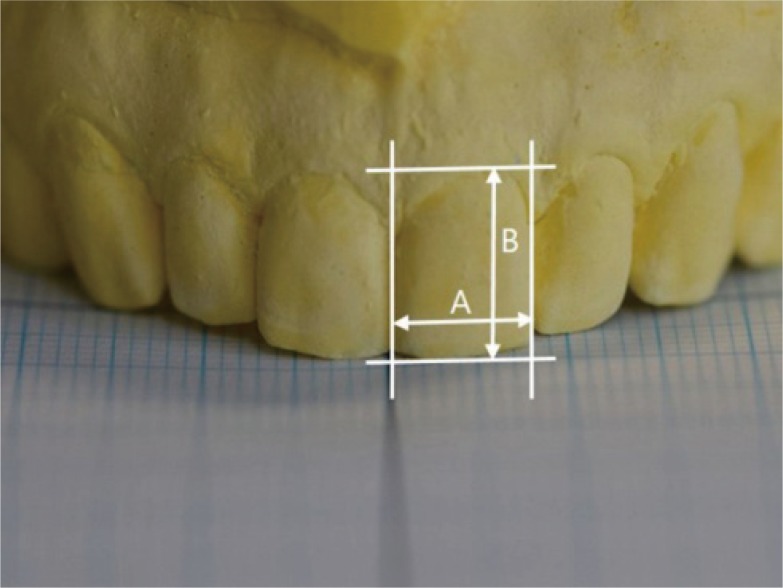
Fig. 3 The visible mesio-distal width (A) and apicocoronal length (B) of maxillary central incisor.
Cited by 1 articles
-
Esthetic restoration of maxillary anterior fixed prosthesis using a digital diagnostic wax-up: a case report
Eunji Oh, Songyi Park, Woohyung Jang, Chan Park, Kwi-Dug Yun, Hyun-Pil Lim, Sangwon Park
J Dent Rehabil Appl Sci. 2023;39(2):89-95. doi: 10.14368/jdras.2023.39.2.89.
Reference
-
1. Goldstein RE. Study of need for esthetics in dentistry. J Prosthet Dent. 1969; 21:589–598. PMID: 5254499.
Article2. Ku JE, Yang HS, Yun KD. A morphometric analysis of maxillary central incisor on the basis of facial appearance in Korea. J Adv Prosthodont. 2012; 4:13–17. PMID: 22439095.
Article3. Lombardi RE. The principles of visual perception and their clinical application to denture esthetics. J Prosthet Dent. 1973; 29:358–382. PMID: 4570911.
Article4. Levin EI. Dental esthetics and the golden proportion. J Prosthet Dent. 1978; 40:244–252. PMID: 279670.
Article5. Ricketts RM. Divine proportion in facial esthetics. Clin Plast Surg. 1982; 9:401–422. PMID: 7172592.
Article6. Preston JD. The golden proportion revisited. J Esthet Dent. 1993; 5:247–251. PMID: 7993669.
Article7. Gillen RJ, Schwartz RS, Hilton TJ, Evans DB. An analysis of selected normative tooth proportions. Int J Prosthodont. 1994; 7:410–417. PMID: 7802908.8. Ali Fayyad M, Jamani KD, Agrabawi J. Geometric and mathematical proportions and their relations to maxillary anterior teeth. J Contemp Dent Pract. 2006; 7:62–70. PMID: 17091141.9. Rosenstiel SF, Ward DH, Rashid RG. Dentists' preferences of anterior tooth proportion-a web-based study. J Prosthodont. 2000; 9:123–136. PMID: 11179463.
Article10. Hasanreisoglu U, Berksun S, Aras K, Arslan I. An analysis of maxillary anterior teeth: facial and dental proportions. J Prosthet Dent. 2005; 94:530–538. PMID: 16316799.
Article11. Sterrett JD, Oliver T, Robinson F, Fortson W, Knaak B, Russell CM. Width/length ratios of normal clinical crowns of the maxillary anterior dentition in man. J Clin Periodontol. 1999; 26:153–157. PMID: 10100040.
Article12. Tsukiyama T, Marcushamer E, Griffin TJ, Arguello E, Magne P, Gallucci GO. Comparison of the anatomic crown width/length ratios of unworn and worn maxillary teeth in Asian and white subjects. J Prosthet Dent. 2012; 107:11–16. PMID: 22230911.
Article13. Ward DH. Proportional smile design using the recurring esthetic dental (red) proportion. Dent Clin North Am. 2001; 45:143–154. PMID: 11210692.14. Jin MX, Hong MH, Lee KJ, Lee KB. Does the maxillary anterior ratio in Korean adults follow the Golden Proportion? J Adv Prosthodont. 2016; 8:125–130. PMID: 27141256.
Article15. Lee SP, Lee SJ, Hayashi K, Park YS. A three-dimensional analysis of the perceived proportions of maxillary anterior teeth. Acta Odontol Scand. 2012; 70:432–440. PMID: 21780976.
Article16. Magne P, Gallucci GO, Belser UC. Anatomic crown width/length ratios of unworn and worn maxillary teeth in white subjects. J Prosthet Dent. 2003; 89:453–461. PMID: 12806322.
Article17. Al-Marzok MI, Majeed KR, Ibrahim IK. Evaluation of maxillary anterior teeth and their relation to the golden proportion in Malaysian population. BMC Oral Health. 2013; 13:9. PMID: 23347800.
Article18. Kook YA, Nojima K, Moon HB, McLaughlin RP, Sinclair PM. Comparison of arch forms between Korean and North American white populations. Am J Orthod Dentofacial Orthop. 2004; 126:680–686. PMID: 15592215.
Article19. Nojima K, McLaughlin RP, Isshiki Y, Sinclair PM. A comparative study of Caucasian and Japanese mandibular clinical arch forms. Angle Orthod. 2001; 71:195–200. PMID: 11407772.20. Schierz O, Dommel S, Hirsch C, Reissmann DR. Occlusal tooth wear in the general population of Germany: effects of age, sex, and location of teeth. J Prosthet Dent. 2014; 112:465–471. PMID: 24636759.
Article21. Yun HJ, Jeong JS, Pang NS, Kwon IK, Jung BY. Radiographic assessment of clinical root-crown ratios of permanent teeth in a healthy Korean population. J Adv Prosthodont. 2014; 6:171–176. PMID: 25006380.
Article22. Peixoto LM, Louro RL, Gomes AA, do Nascimento APC. Photographic analysis of esthetic dental proportions. Rev Gaucha Odontol. 2012; 60:13–17.23. Cooper GE, Tredwin CJ, Cooper NT, Petrie A, Gill DS. The influence of maxillary central incisor height-to-width ratio on perceived smile aesthetics. Br Dent J. 2012; 212:589–599. PMID: 22722122.
Article24. Hattab FN, Yassin OM. Etiology and diagnosis of tooth wear: a literature review and presentation of selected cases. Int J Prosthodont. 2000; 13:101–107. PMID: 11203616.
- Full Text Links
-
- Actions
-
Cited
- CITED
-
- Close
- Share
-
- Similar articles
-
- The effect of mesiodistal crown widths of anterior teeth on incisor relationship
- Golden proportion assessment between maxillary and mandibular teeth on Indian population
- Study of Normative Gingival Proportion in Anterior Maxilla
- Fourier Analysis of Maxillary Dental Arch Forms
- Analysis of esthetic factors and evaluation of esthetic perception for maxillary anteriors of dental students
