Extraosseous Ewing's Sarcoma Presented as a Rectal Subepithelial Tumor: Radiological and Pathological Features
- Affiliations
-
- 1Department of Radiology, Yonsei University College of Medicine, Seoul, Korea. pine0205@yuhs.ac
- 2Research Institute of Radiological Science, Yonsei University College of Medicine, Seoul, Korea.
- 3Department of Pathology, Yonsei University College of Medicine, Seoul, Korea.
- KMID: 2376270
- DOI: http://doi.org/10.13104/imri.2017.21.1.51
Abstract
- PURPOSE
Extraosseous Ewing's sarcoma (EOE) of the rectum is extremely rare: only three cases have been reported in the literature and none of these reports described their imaging findings in detail. Herein, we describe the tumor imaging and pathological features in detail.
MATERIALS AND METHODS
We report a case of rectal EOE in a 72-year-old female who received local excision and was provisionally diagnosed with a rectal submucosal spindle cell tumor. We used immunohistochemistry, histopathology, and fluorescence in situ hybridization to characterize the tumor and provide a definitive diagnosis of EOE.
RESULTS
MRI revealed a well-demarcated submucosal tumor with heterogeneous enhancement and hemorrhagic foci in rectum. EOE was diagnosed by positive staining of tumor cells for CD99 and Fli-1 by immunohistochemistry and the presence of the EWSR1 gene translocation by fluorescence in situ hybridization. Although the patient underwent radiation treatment and surgery, the tumor recurred after 4 months as revealed by computed tomography and magnetic resonance imaging.
CONCLUSION
Rectal EOE may present as a rectal submucosal tumor. The understanding of imaging and histological characteristics of this tumor are critical for accurate diagnosis and appropriate aggressive treatment.
MeSH Terms
Figure
-
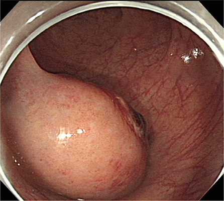
Fig. 1 Initial colonoscopy image demonstrating a rectal subepithelial tumor.

Fig. 2 Rectal MRI: (a) Precontrast T1-weighted axial image, (b) T2-weighted oblique axial image, and (c) contrast-enhanced T1-weighted axial and (d) contrast-enhanced T1-weighted coronal image revealed a well-defined submucosal mass measuring approximately 4 cm in the lower rectum. The mass (arrows) showed heterogeneous enhancement with a focal hemorrhage (arrowheads).
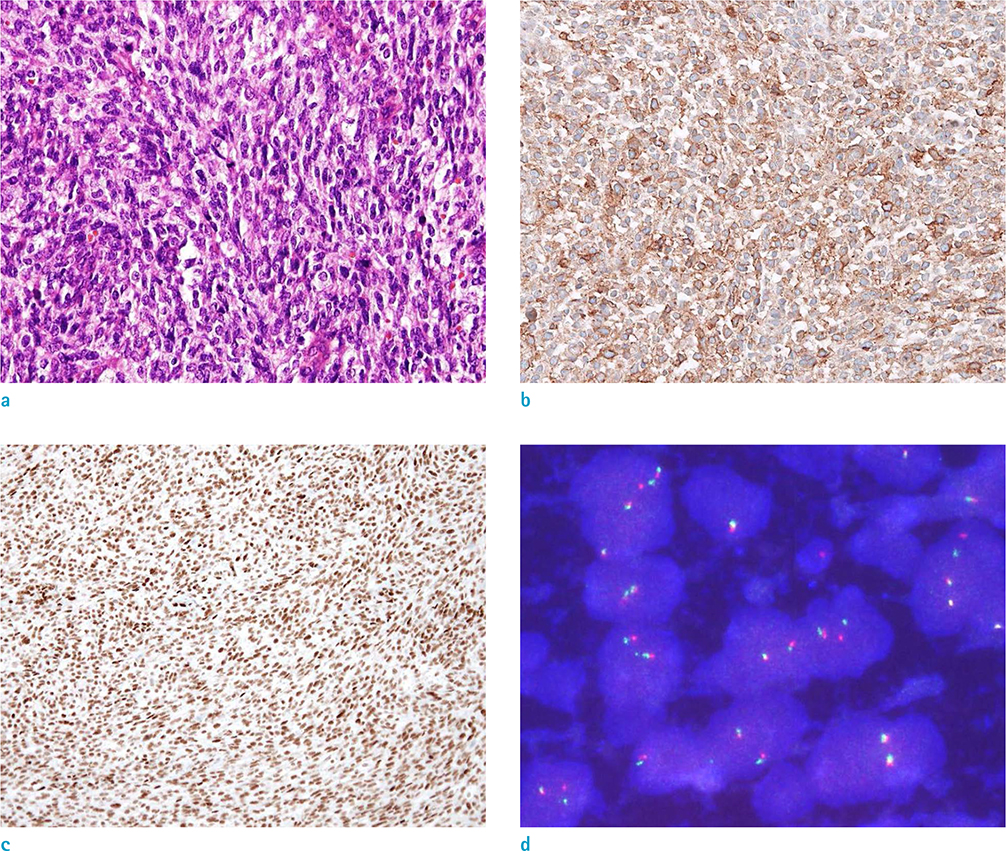
Fig. 3 Microscopic examination of the local excised specimen. (a) Hematoxylin and Eosin stain (× 200). Immunohistochemistry of (b) CD99 (× 200) and (c) Fli-1 (× 100). (d) Fluorescence in situ hybridization demonstrated the EWSR1 gene translocation, which confirmed the pathological diagnosis of Ewing's sarcoma (× 1000).

Fig. 4 Preoperative imaging studies of the locally recurrent mass. Comparison of (a) CT obtained when the locally recurrent mass (arrow) was detected, and (b, c) MRI obtained 2 months after pelvic irradiation indicated that the size of the mass had partially decreased after irradiation. (b) Upon T2-weighted oblique axial imaging, markedly low T2 signal intensity (black arrow) of the residual lesion suggested that extensive fibrotic change had occurred. (c) Contrast enhanced T1-weighted axial image showed minimal enhancement (white arrow) within the residual lesion.
Reference
-
1. Fletcher CD, Bridge JA, Hogendoorn P, Mertens F. WHO classification of tumours of soft tissue and bone. 4th ed. Lyon, France: International Agency for Research on Cancer;2013.2. Vardy J, Joshua AM, Clarke SJ, Yarrow PM, Lin BP. Small blue cell tumors of the rectum. Case 1. Ewing's sarcoma of the rectum. J Clin Oncol. 2005; 23:910–912.3. Aboumarzouk OM, Coleman R, Goepel JR, Shorthouse AJ. PNET/Ewing's sarcoma of the rectum: a case report and review of the literature. BMJ Case Rep. 2009; 2009:pii: bcr04.2009.1770.4. Javery O, Krajewski K, O'Regan K, et al. A to Z of extra-skeletal Ewing sarcoma family of tumors in adults: imaging features of primary disease, metastatic patterns, and treatment responses. AJR Am J Roentgenol. 2011; 197:W1015–W1022.5. Kim SK, Park YK. Ewing sarcoma: a chronicle of molecular pathogenesis. Hum Pathol. 2016; 55:91–100.6. Wei S, Siegal GP. Round cell tumors of bone: an update on recent molecular genetic advances. Adv Anat Pathol. 2014; 21:359–372.7. Murphey MD, Senchak LT, Mambalam PK, Logie CI, Klassen-Fischer MK, Kransdorf MJ. From the radiologic pathology archives: ewing sarcoma family of tumors: radiologic-pathologic correlation. Radiographics. 2013; 33:803–831.8. Huh J, Kim KW, Park SJ, et al. Imaging features of primary tumors and metastatic patterns of the extraskeletal ewing sarcoma family of tumors in adults: a 17-year experience at a single institution. Korean J Radiol. 2015; 16:783–790.9. Kim H, Kim JH, Lim JS, et al. MRI findings of rectal submucosal tumors. Korean J Radiol. 2011; 12:487–498.10. Angervall L, Enzinger FM. Extraskeletal neoplasm resembling Ewing's sarcoma. Cancer. 1975; 36:240–251.
