Investig Magn Reson Imaging.
2017 Mar;21(1):28-33. 10.13104/imri.2017.21.1.28.
Measurement and Compensation of Respiration-Induced B0 Variations in Lumbar Spine Bone Marrow Fat Quantification
- Affiliations
-
- 1Department of Radiology, Seoul St. Mary's Hospital, College of Medicine, The Catholic University of Korea, Seoul, Korea. jjdragon112@gmail.com
- KMID: 2376266
- DOI: http://doi.org/10.13104/imri.2017.21.1.28
Abstract
- PURPOSE
To investigate and compensate the effects of respiration-induced B0 variations on fat quantification of the bone marrow in the lumbar spine.
MATERIALS AND METHODS
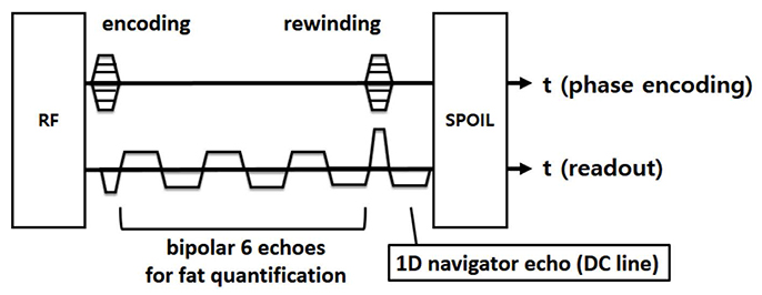
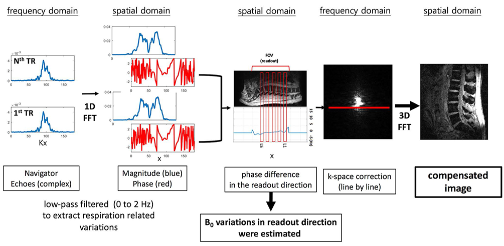
Multi-echo gradient echo images with navigator echoes were obtained from eight healthy volunteers at 3T clinical scanner. Using navigator echo data, respiration-induced B0 variations were measured and compensated. Fat fraction maps were estimated using T2*-IDEAL algorithm from the uncompensated and compensated images. For manually drawn bone marrow regions, the estimated B0 variations and the calculated fat fractions (before and after compensations) were analyzed.
RESULTS
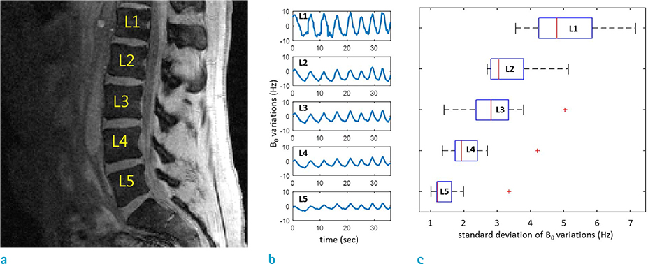
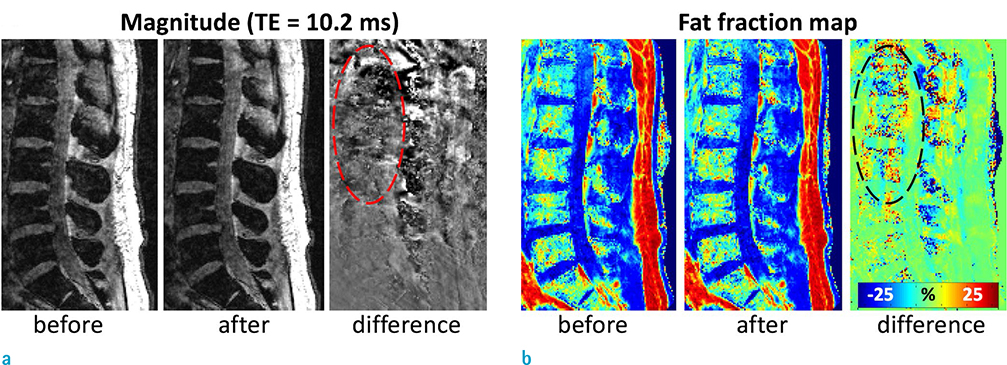
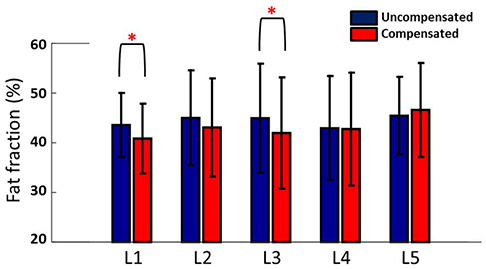
An increase of temporal B0 variations from inferior level to superior levels was observed for all subjects. After compensation using navigator echo data, the effects of the B0 variations were reduced in gradient echo images. The calculated fat fractions show significant differences (P < 0.05) in L1 and L3 between the uncompensated and the compensated.
CONCLUSION
The results of this study raise the need for considering respiration-induced B0 variations for accurate fat quantification using gradient echo images in the lumbar spine. The use of navigator echo data can be an effective way for the reduction of the effects of respiratory motion on the quantification.
Keyword
Figure
Reference
-
1. Ragab Y, Emad Y, Gheita T, et al. Differentiation of osteoporotic and neoplastic vertebral fractures by chemical shift {in-phase and out-of phase} MR imaging. Eur J Radiol. 2009; 72:125–133.2. Padhani AR, Makris A, Gall P, Collins DJ, Tunariu N, de Bono JS. Therapy monitoring of skeletal metastases with whole-body diffusion MRI. J Magn Reson Imaging. 2014; 39:1049–1078.3. Baum T, Yap SP, Dieckmeyer M, et al. Assessment of whole spine vertebral bone marrow fat using chemical shift-encoding based water-fat MRI. J Magn Reson Imaging. 2015; 42:1018–1023.4. Li X, Kuo D, Schafer AL, et al. Quantification of vertebral bone marrow fat content using 3 Tesla MR spectroscopy: reproducibility, vertebral variation, and applications in osteoporosis. J Magn Reson Imaging. 2011; 33:974–979.5. Rosen CJ, Ackert-Bicknell C, Rodriguez JP, Pino AM. Marrow fat and the bone microenvironment: developmental, functional, and pathological implications. Crit Rev Eukaryot Gene Expr. 2009; 19:109–124.6. Schellinger D, Lin CS, Fertikh D, et al. Normal lumbar vertebrae: anatomic, age, and sex variance in subjects at proton MR spectroscopy--initial experience. Radiology. 2000; 215:910–916.7. Ma J. Dixon techniques for water and fat imaging. J Magn Reson Imaging. 2008; 28:543–558.8. Yu H, McKenzie CA, Shimakawa A, et al. Multiecho reconstruction for simultaneous water-fat decomposition and T2* estimation. J Magn Reson Imaging. 2007; 26:1153–1161.9. Verma T, Cohen-Adad J. Effect of respiration on the B0 field in the human spinal cord at 3T. Magn Reson Med. 2014; 72:1629–1636.10. Yu H, Shimakawa A, McKenzie CA, et al. Phase and amplitude correction for multi-echo water-fat separation with bipolar acquisitions. J Magn Reson Imaging. 2010; 31:1264–1271.11. Hu X, Kim SG. Reduction of signal fluctuation in functional MRI using navigator echoes. Magn Reson Med. 1994; 31:495–503.12. Wen J, Cross AH, Yablonskiy DA. On the role of physiological fluctuations in quantitative gradient echo MRI: implications for GEPCI, QSM, and SWI. Magn Reson Med. 2015; 73:195–203.13. Nam Y, Kim DH, Lee J. Physiological noise compensation in gradient-echo myelin water imaging. Neuroimage. 2015; 120:345–349.14. Yoo YH, Kim HS, Lee YH, et al. Comparison of Multi-Echo Dixon Methods with Volume Interpolated Breath-Hold Gradient Echo Magnetic Resonance Imaging in Fat-Signal Fraction Quantification of Paravertebral Muscle. Korean J Radiol. 2015; 16:1086–1095.15. Kang BK, Yu ES, Lee SS, et al. Hepatic fat quantification: a prospective comparison of magnetic resonance spectroscopy and analysis methods for chemical-shift gradient echo magnetic resonance imaging with histologic assessment as the reference standard. Invest Radiol. 2012; 47:368–375.16. Yokoo T, Shiehmorteza M, Hamilton G, et al. Estimation of hepatic proton-density fat fraction by using MR imaging at 3.0 T. Radiology. 2011; 258:749–759.17. Lee H, Nam Y, Han D, Gho SM, Kim DH. SWI of the cervical-spinal cord with respiration noise correction using navigator echo. In : ISMRM 23rd Annual Meeting & Exhibition; 30 May-5 June, 2015; Toronto, Ontario, Canada. #1727.
- Full Text Links
- Actions
-
Cited
- CITED
-
- Close
- Share
- Similar articles
-
- Background Gradient Correction using Excitation Pulse Profile for Fat and T2* Quantification in 2D Multi-Slice Liver Imaging
- High-Resolution Numerical Simulation of Respiration-Induced Dynamic Bâ‚€ Shift in the Head in High-Field MRI
- Susceptibility Weighted Imaging of the Cervical Spinal Cord with Compensation of Respiratory-Induced Artifact
- Quantifying Bone Marrow Edema Adjacent to the Lumbar Vertebral Endplate on Magnetic Resonance Imaging: A Cross-Sectional Study of Patients with Degenerative Lumbar Disease
- Rapid Increase in Marrow Fat Content and Decrease in Marrow Perfusion in Lumbar Vertebra Following Bilateral Oophorectomy: An MR Imaging-Based Prospective Longitudinal Study

