Korean J Radiol.
2015 Feb;16(1):154-159. 10.3348/kjr.2015.16.1.154.
Rapid Increase in Marrow Fat Content and Decrease in Marrow Perfusion in Lumbar Vertebra Following Bilateral Oophorectomy: An MR Imaging-Based Prospective Longitudinal Study
- Affiliations
-
- 1Department of Imaging & Interventional Radiology, Prince of Wales Hospital, The Chinese University of Hong Kong, Shatin, N.T., Hong Kong SAR, China. yixiang_wang@cuhk.edu.hk
- KMID: 2069994
- DOI: http://doi.org/10.3348/kjr.2015.16.1.154
Abstract
OBJECTIVE
Bilateral oophorectomy leads to reduced bone mineral density (BMD), and reduced BMD is associated with increased marrow fat and reduced marrow perfusion. Purpose of this study was to investigate how soon these changes occur following surgical oophorectomy.
MATERIALS AND METHODS
Six patients who underwent hysterectomy and bilateral salpingo-oophorectomy were studied. At baseline, mean patient age was 49.5 years (range: 45-54 years). Third lumbar vertebral body BMD measurement using quantitative CT, marrow fat fraction (FF) using MR spectroscopy and marrow perfusion using dynamic contrast enhanced MRI were conducted immediately prior to surgery and at 3, 9, and 21 months after surgery.
RESULTS
Reduced BMD, increased marrow FF, and reduced marrow perfusion occurred synchronously post-oophorectomy. There was a sharp decrease of 12.5 +/- 7.2% in BMD (n = 6), a sharp increase of 92.2 +/- 46.3% (n = 6) in FF, a sharp decrease of 23.6 +/- 3.9% in maximum contrast enhancement (n = 5), and of 45.4 +/- 7.7% for enhancement slope (n = 5) during the initial 3 months post surgery. BMD and marrow perfusion continued to decrease, and marrow FF continued to increase at a slower rate during the following 18 months. Friedman test showed a significant trend for these changes (p < 0.05).
CONCLUSION
Bilateral oophorectomy leads to a rapid decrease in lumbar BMD, an increase in marrow fat content, and a decrease in marrow blood perfusion.
Keyword
MeSH Terms
Figure
-
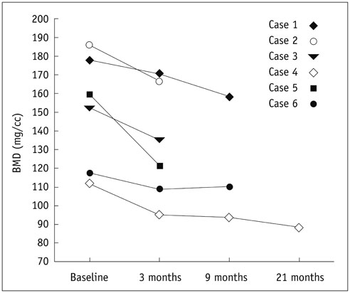
Fig. 1 Lumbar vertebral bone mineral density (BMD) during course of study.
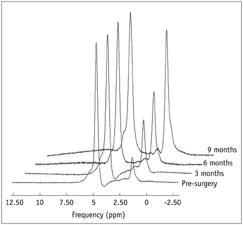
Fig. 2 Typical MR spectra presentation of marrow fat fraction increase post oophorectomy. Water peak is at 4.65 ppm and lipid peak is at 1.3 ppm.
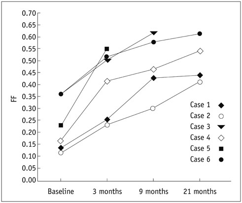
Fig. 3 Fat fraction (FF) changes during course of study.
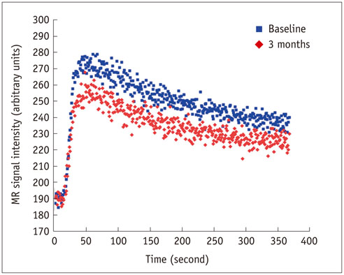
Fig. 4 Typical time-intensity enhancement curve at baseline (top trace) and 3 months post oophorectomy (bottom trace). Time-intensity curve post surgery shows reduced Emax and less steep Eslope.
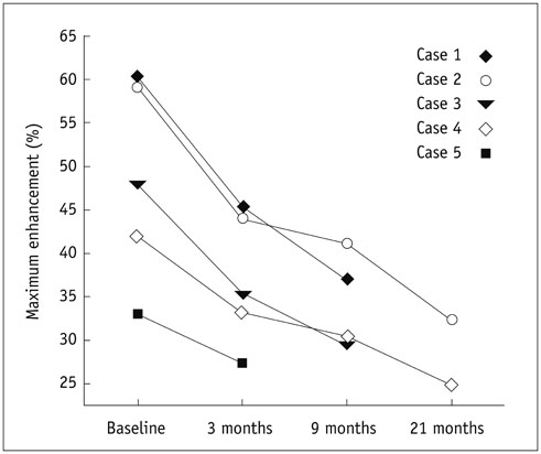
Fig. 5 Maximum enhancement (Emax) during course of study.
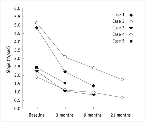
Fig. 6 Enhancement slope (Eslope) during course of study.
Reference
-
1. Gallagher JC. Effect of early menopause on bone mineral density and fractures. Menopause. 2007; 14(3 Pt 2):567–571.2. Deng M, Wang YX, Griffith JF, Lu G, Ahuja AT, Poon WS. Characteristics of rat lumbar vertebral body bone mineral density and differential segmental responses to sex hormone deficiency: a clinical multidetector computed tomography study. Biomed Environ Sci. 2012; 25:607–613.3. Bellino FL, Wise PM. Nonhuman primate models of menopause workshop. Biol Reprod. 2003; 68:10–18.4. Deng M, Griffith JF, Zhu XM, Poon WS, Ahuja AT, Wang YX. Effect of ovariectomy on contrast agent diffusion into lumbar intervertebral disc: a dynamic contrast-enhanced MRI study in female rats. Magn Reson Imaging. 2012; 30:683–688.5. Davies MC, Hall ML, Jacobs HS. Bone mineral loss in young women with amenorrhoea. BMJ. 1990; 301:790–793.6. Cann CE, Martin MC, Genant HK, Jaffe RB. Decreased spinal mineral content in amenorrheic women. JAMA. 1984; 251:626–629.7. Wolman RL, Clark P, McNally E, Harries M, Reeve J. Menstrual state and exercise as determinants of spinal trabecular bone density in female athletes. BMJ. 1990; 301:516–518.8. Shih TT, Liu HC, Chang CJ, Wei SY, Shen LC, Yang PC. Correlation of MR lumbar spine bone marrow perfusion with bone mineral density in female subjects. Radiology. 2004; 233:121–128.9. Griffith JF, Yeung DK, Antonio GE, Lee FK, Hong AW, Wong SY, et al. Vertebral bone mineral density, marrow perfusion, and fat content in healthy men and men with osteoporosis: dynamic contrast-enhanced MR imaging and MR spectroscopy. Radiology. 2005; 236:945–951.10. Griffith JF, Yeung DK, Antonio GE, Wong SY, Kwok TC, Woo J, et al. Vertebral marrow fat content and diffusion and perfusion indexes in women with varying bone density: MR evaluation. Radiology. 2006; 241:831–838.11. Wang YX, Griffith JF, Kwok AW, Leung JC, Yeung DK, Ahuja AT, et al. Reduced bone perfusion in proximal femur of subjects with decreased bone mineral density preferentially affects the femoral neck. Bone. 2009; 45:711–715.12. Wang YX, Zhang YF, Griffith JF, Zhou H, Yeung DK, Kwok TC, et al. Vertebral blood perfusion reduction associated with vertebral bone mineral density reduction: a dynamic contrast-enhanced MRI study in a rat orchiectomy model. J Magn Reson Imaging. 2008; 28:1515–1518.13. Griffith JF, Yeung DK, Ma HT, Leung JC, Kwok TC, Leung PC. Bone marrow fat content in the elderly: a reversal of sex difference seen in younger subjects. J Magn Reson Imaging. 2012; 36:225–230.14. Sanada M, Taguchi A, Higashi Y, Tsuda M, Kodama I, Yoshizumi M, et al. Forearm endothelial function and bone mineral loss in postmenopausal women. Atherosclerosis. 2004; 176:387–392.15. Sumino H, Ichikawa S, Kasama S, Takahashi T, Sakamoto H, Kumakura H, et al. Relationship between brachial arterial endothelial function and lumbar spine bone mineral density in postmenopausal women. Circ J. 2007; 71:1555–1559.16. Williams JK, Honoré EK, Washburn SA, Clarkson TB. Effects of hormone replacement therapy on reactivity of atherosclerotic coronary arteries in cynomolgus monkeys. J Am Coll Cardiol. 1994; 24:1757–1761.17. Wang YX, Zhou H, Griffith JF, Zhang YF, Yeung DK, Ahuja AT. An in vivo magnetic resonance imaging technique for measurement of rat lumbar vertebral body blood perfusion. Lab Anim. 2009; 43:261–265.18. Zhang YF, Wang YX, Griffith JF, Kwong WK, Ma HT, Qin L, et al. Proximal femur bone marrow blood perfusion indices are reduced in hypertensive rats: a dynamic contrast-enhanced MRI study. J Magn Reson Imaging. 2009; 30:1139–1144.19. Griffith JF, Yeung DK, Chow SK, Leung JC, Leung PC. Reproducibility of MR perfusion and (1)H spectroscopy of bone marrow. J Magn Reson Imaging. 2009; 29:1438–1442.20. Pansini F, Bagni B, Bonaccorsi G, Albertazzi P, Zanotti L, Farina A, et al. Oophorectomy and Spine Bone Density: Evidence of a Higher Rate of Bone Loss in Surgical Compared with Spontaneous Menopause. Menopause. 1995; 2:109–115.21. Prior JC, Vigna YM, Wark JD, Eyre DR, Lentle BC, Li DK, et al. Premenopausal ovariectomy-related bone loss: a randomized, double-blind, one-year trial of conjugated estrogen or medroxyprogesterone acetate. J Bone Miner Res. 1997; 12:1851–1863.22. Griffith JF, Wang YX, Zhou H, Kwong WH, Wong WT, Sun YL, et al. Reduced bone perfusion in osteoporosis: likely causes in an ovariectomy rat model. Radiology. 2010; 254:739–746.23. Basu S, Houseni M, Bural G, Chamroonat W, Udupa J, Mishra S, et al. Magnetic resonance imaging based bone marrow segmentation for quantitative calculation of pure red marrow metabolism using 2-deoxy-2-[F-18]fluoro-D-glucose-positron emission tomography: a novel application with significant implications for combined structure-function approach. Mol Imaging Biol. 2007; 9:361–365.24. Chen WT, Shih TT, Chen RC, Lo SY, Chou CT, Lee JM, et al. Vertebral bone marrow perfusion evaluated with dynamic contrast-enhanced MR imaging: significance of aging and sex. Radiology. 2001; 220:213–218.25. Kita K, Kawai K, Hirohata K. Changes in bone marrow blood flow with aging. J Orthop Res. 1987; 5:569–575.26. Hartsock RJ, Smith EB, Petty CS. Normal variations with aging of the amount of hematopoietic tissue in bone marrow from the anterior iliac crest. A study made from 177 cases of sudden death examined by necropsy. Am J Clin Pathol. 1965; 43:326–331.27. Ito H. Clinical considerations of regenerative medicine in osteoporosis. Curr Osteoporos Rep. 2014; 12:230–234.28. Wang YX, Ng CK. The impact of quantitative imaging in medicine and surgery: charting our course for the future. Quant Imaging Med Surg. 2011; 1:1–3.29. Masarapu V, Kim HL. Initial experience with arterial spin-labeling MR imaging to assess histology of renal masses. Quant Imaging Med Surg. 2013; 3:130–131.30. Martirosian P, Boss A, Schraml C, Schwenzer NF, Graf H, Claussen CD, et al. Magnetic resonance perfusion imaging without contrast media. Eur J Nucl Med Mol Imaging. 2010; 37:Suppl 1. S52–S64.31. Roldan-Valadez E, Piña-Jimenez C, Favila R, Rios C. Gender and age groups interactions in the quantification of bone marrow fat content in lumbar spine using 3T MR spectroscopy: a multivariate analysis of covariance (Mancova). Eur J Radiol. 2013; 82:e697–e702.
- Full Text Links
-
- Actions
-
Cited
- CITED
-
- Close
- Share
-
- Similar articles
-
- Age- Related Contrast Enhancement Study of Normal Bone Marrow in Lumbar Spinal MR Imaging
- Measurement and Compensation of Respiration-Induced B0 Variations in Lumbar Spine Bone Marrow Fat Quantification
- Magnetic Resonance Imaging of the Bone Marrow After Bone Marrow Transplantation or Immunosuppressive Therapy in Aplastic Anemia
- Assessement of MR signal intensity of cranium and cervical spine bone marrow
- MR Imaging Findings of Bone Marrow Following Bone Marrow Transplantation
