Synchrotron X-ray Microscopic Imaging of Thyroid Tissues
- Affiliations
-
- 1Department of Surgery, College of Medicine, Catholic University of Daegu, Daegu, Korea. shwpark@cu.ac.kr
- 2Pohang Accelerator Laboratory, Pohang University of Science and Technology, Pohang, Korea.
- KMID: 2375728
- DOI: http://doi.org/10.16956/kjes.2010.10.1.19
Abstract
- PURPOSE
X-ray microscopy with synchrotron radiation will soon be a useful tool for innovative x-ray imaging in clinical and laboratory settings. It enables us to observe the detailed internal structure of human tissue samples with great magnification power and excellent resolution. So, it has the possibility to be used for the clinical and research purposes to investigate thyroid diseases if it can effectively evaluate the various conditions of thyroid tissue. To determine the relation with their optical microscopic features, we compared the synchrotron X-ray images of unstained normal and thyroid cancer tissue samples with the histopathologic findings of their adjacent, stained thyroid tissue sections.
METHODS
An x-ray microscope was installed on a 1B2 beamline with a Pohang Light Source, which is a 3rd generation synchrotron radiation facility with an operating energy of 2.5 GeV at Pohang, Korea. The x-ray energy was set at 11.1 keV and the x-ray beam was monochromatized using a W/B4C monochromator. Formalin-fixed 10µm-thick female thyroid tissues from normal cases and carcinoma cases were attached on Kapton film for the imaging. The sample was positioned 25 m away from the beam source. The x-ray image of the sample was converted into a visual image on the CsI (TI) scintillation crystal, and it was magnified 20 times by the microscopic objective lens. After an additional 10 times digital magnification, this visual image was captured by a full frame CCD camera.
RESULTS
The monochromated x-ray microscopic images of the female thyroid tissues of the normal cases and carcinoma cases were obtained with good resolution. These synchrotron images showed the normal follicular structures in the normal thyroid tissue sections and the characteristic severe stromal fibrosis with collagen fiber accumulation in the cancer tissue sections.
CONCLUSION
Owing to the great magnification and excellent resolution, the synchrotron x-ray microscopic images of the normal and cancerous thyroid tissues showed good correspondence with the histopathologic findings of their adjacent, stained tissue sections. So, the x-ray microscopic imaging of thyoid tissue using synchrotron radiation has good potential for use in various clinical and research settings in the future.
Keyword
MeSH Terms
Figure
-
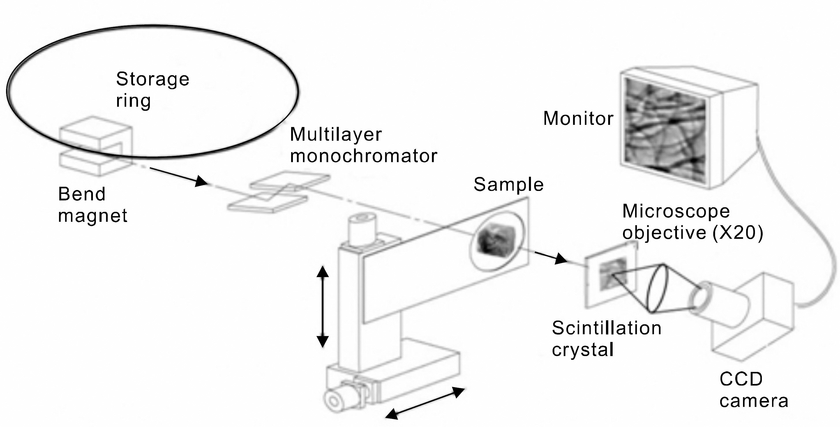
Fig. 1 This is the layout of the 1B2 beamline optics for an x-ray microscope at Pohang Light Source in Pohang, Korea.
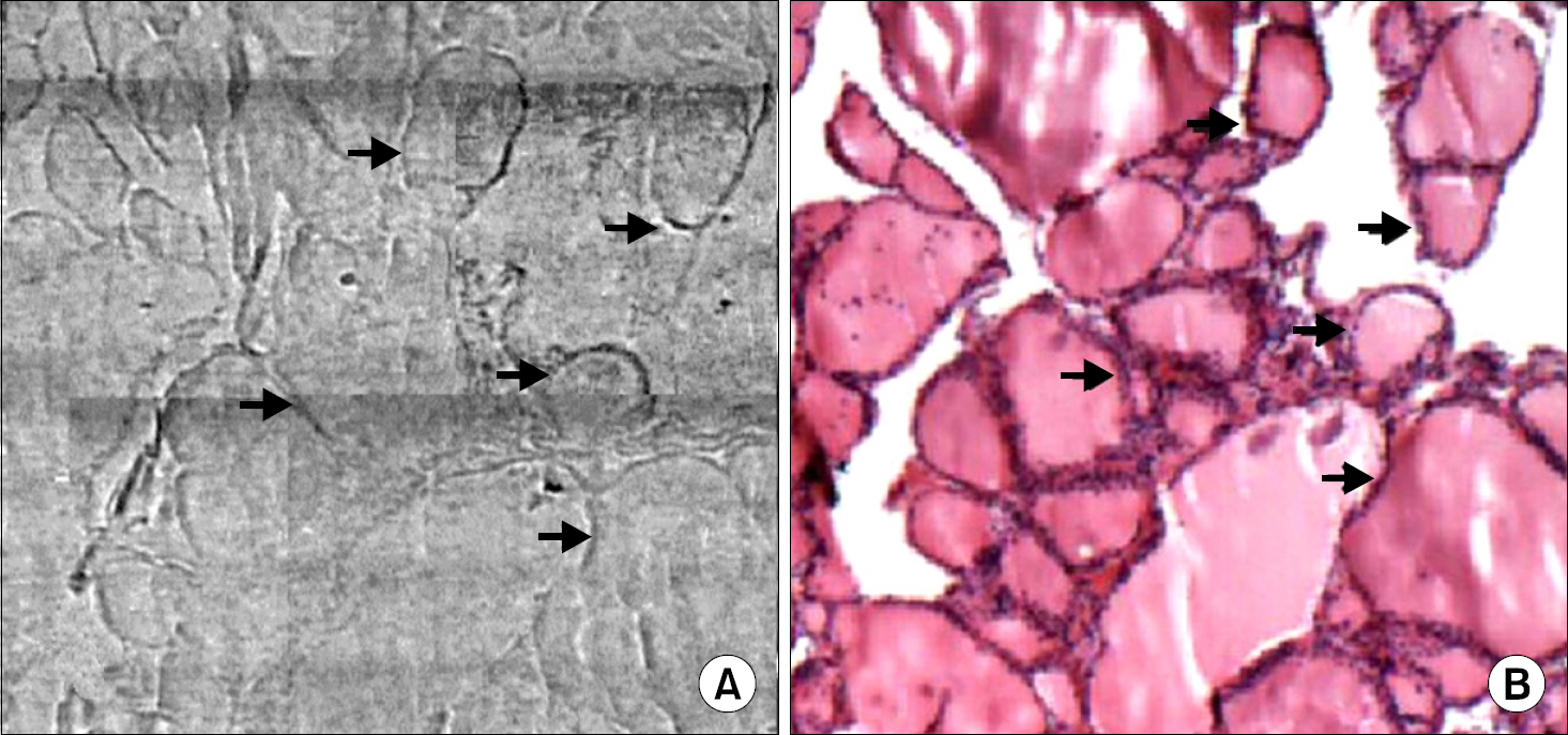
Fig. 2 Synchrotron image (A) and histologic section (B) of normal thyroid tissue. Synch-rontron image shows distinctly outlined colloid filled thyroid follicles by thin radio-opaque line (arrows) of follicular cells and basement membrane, which well correlated to the histologic section (H&E, ×200).

Fig. 3 Comparison of synchrotron image (A) and histologic section (B) of papillary carcinoma of the thyroid. The synchrotron image is well correlated to the histologic section with prominent peritumoral fibrosis. The infiltrating tumor cell nests (∗) are surrounded by thick collagenous fibers (arrows), which are highlighted in the synchrotron image (H&E, ×100).
Reference
-
1.Youn HS., Jung SW. Observations of a human hair shaft with an x-ray microscope. Phys Med Biol. 2005. 50:5417–20.
Article2.Choi CH., Kim HT., Choe JY., Kim JK., Youn HS. Application of synchrotron radiation imaging for non-destructive monitoring of mouse rheumatoid arthritis model. American Institute of Physics. 2007. 879:1952–5.
Article3.Jheon SH., Youn HS., Kim HT., Kim JK. High-resolution x-ray refraction imaging of rat lung and histological correlations. Microsc Res Tech. 2006. 69:656–9.
Article4.Arfelli F. Synchrotron light and imaging systems for medical radiology. Nucl Instrum Meth Phys Res A. 2000. 454:11–25.
Article5.Jeong YJ., Bong JG., Kim HT., Kim JK., Jheon SH., Youn HS, et al. Synchrotron radiation imaging of female breast tissues using phase contrast technique. J Breast Cancer. 2008. 11:40–4.
Article6.Arfelli F., Assante M., Bonvicini V., Bravin A., Cantatore G., Castelli E, et al. Low dose phase contrast x-ray medical imaging. Phys Med Biol. 1998. 43:2845–52.7.Park SH., Kim HT., Kim JK., Jheon SH., Youn HS. Hard x-ray microscopic imaging of human breast tissues. AIP Conf Proc. 2007. 879:1976–8.
Article8.Rochaa H., Lopesa R., Valianteb P., Tiraoc G., Mazzarod I., Honnicked M, et al. Diagnosis of thyroid multinodular goiter using diffraction-enhanced imaging. Nucl Instrum Meth Phys Res A. 2005. 548:175–80.9.Ando M., Bando H., Chen Z., Chikaura Y., Choi CH., Endo T, et al. 2D and 3D refraction based x-ray imaging suitable for clinical and pathological diagnosis. American Institute of Physics. 2007. 879:1899–902.
Article10.Hwu Y., Hsieh H., Lu MJ., Tsai WL., Lin HM., Goh WC, et al. Coherence-enhanced synchrotron radiology: Refraction versus diffraction mechanisms. J Appl Phys. 1999. 86:4613–8.
Article11.Takeda T., Yu Q., Yashiro T., Zeniya T., Wu J., Hasegawa T, et al. Iodine imaging in thyroid by fluorescent X-ray CT with 0.05mm spatial resolution. Nucl Instrum Meth Phys Res A. 2001. 467-468:1318–21.
Article12.Takeda T., Zeniya T., Wu J., Yu Q., Lwin T., Tsuchiya Y, et al. Medical Imaging by fluorescent X-ray CT: Its preliminary clinical evaluation. Proc of SPIE. 2002. 4503:299–309.
Article13.Son S., Park S., Park G., Ha S., Lee G., Lee O, et al. Ex Vovo imaging of basal cell carcinoma using synchrotron phase-contrast X-ray microscopy. Skin Res Technol. 2008. 14:13–7.
- Full Text Links
-
- Actions
-
Cited
- CITED
-
- Close
- Share
-
- Similar articles
-
- Synchrotron Radiation Imaging of Breast Tissue Using a Phase-contrast Hard X-ray Microscope
- Synchrotron Radiation Imaging of Female Breast Tissues Using Phase Contrast Technique
- Phase Contrast Microradiography of Mouse Lung Using Synchrotron X-ray: Correlation with Optical Microscopy
- 3D Histology Using the Synchrotron Radiation Propagation Phase Contrast Cryo-microCT
- A Case of microscopic pulmonary metastasis of papillary thyroid carcinoma
