Brown Tumor of the Humerus Associated with Secondary Hyperparathyroidism: A Case Report of Successful Treatment after Subtotal Parathyroidectomy
- Affiliations
-
- 1Department of Otolaryngology-Head and Neck Surgery, Dong-A University College of Medicine, Busan, Korea. hspark1@dau.ac.kr
- KMID: 2375421
- DOI: http://doi.org/10.0000/kjes.2013.13.2.92
Abstract
- Brown tumor is a bone disease that arises in the setting of increased osteoclastic activity and fibroblastic proliferation in the involved bone. It is well recognized as serious complication of hyperparathyroidism. Brown tumor is uncommon, and brown tumor with secondary hyperparathyroidism resulting from chronic renal failure has rarely been reported. We recently experienced a case of a 28-year-old Korean woman with chronic renal failure caused by chronic glomerulonephritis, on hemodialysis for nine years. She has been hospitalized with left shoulder pain for two years. Image studies showed multiple cystic masses, and both suspicious marked thinning and partial destruction of the cortex on the head of the left humerus. Histopathologic analysis of the mass lesion showed a fibrotic capsule, hemosiderin pigmentation, and giant cell, all characteristic of brown tumor. A subtotal parathyroidectomy was done without surgery of the bony lesion (brown tumor), with successful results. We report this case with a brief review of the literature.
MeSH Terms
Figure
-

Fig. 1. Radiologic findings. Simple X-ray: Geographic expansile intramedullary osteolytic lesion from left proximal humerus head portion to metadiaphysis (A). Upper extremity CT axial view: Multiseptated cystic bone tumor in left humerus metadiaphysis area. Suspicious marked thinning and partial destruction in cortex. Edematous change in muscles and soft tissue area (B). MRI shoulder axial view: Multiseptated cystic bone tumor in left humerus metadiaphysis, prominent thinning in cortex, suspicious cortical destruction, edematous change in muscles and soft tissue area (C). Simple X-ray (5 months after surgery): Reduced expansile and osteolytic lesion of the left proximal humerus head portion (D). Upper extremity CT (5 months after surgery): Markedly reduced cystic portion of left humerus (E).
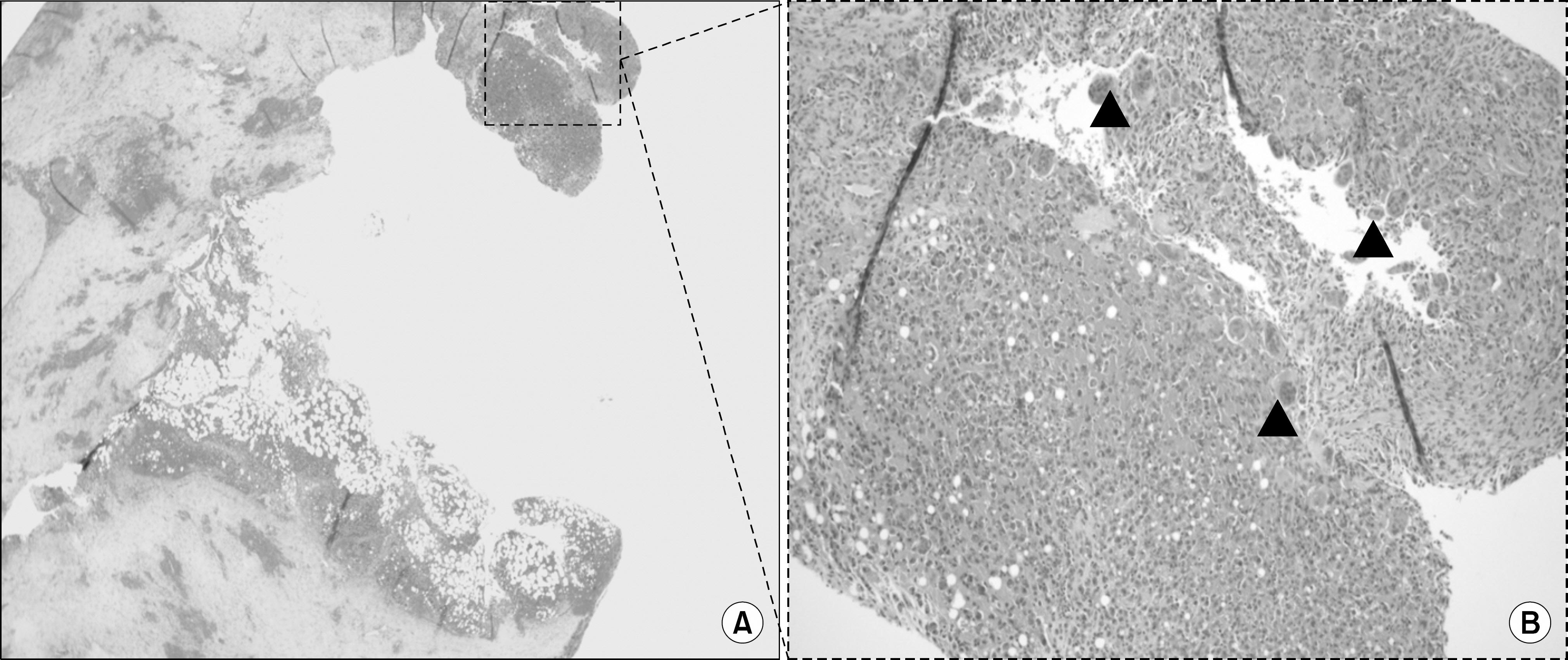
Fig. 2. Pathologic findings. Benign spindle cells and giant cell proliferation lesion with cystic change and hemorrhage (A, ×200), (B, ×400: black arrow heads).

Fig. 3. Neck CT: Low density nodular lesion in posterior portion left thyroid gland (arrow).
Reference
-
References
1. Jeren-Strujić B, Rozman B, Lambasa S, Jeren T, Marković M, Raos V. Secondary hyperparathyroidism and brown tumor in dialyzed patients. Ren Fail. 2001; 23:279–86.
Article2. Yamazaki H, Ota Y, Aoki T, Karakida K. Brown tumor of the maxilla and mandible: progressive mandibular brown tumor after removal of parathyroid adenoma. J Oral Maxillofac Surg. 2003; 61:719–22.
Article3. Fineman I, Johnson JP, Di-Patre PL, Sandhu H. Chronic renal failure causing brown tumors and myelopathy. Case report and review of pathophysiology and treatment. J Neurosurg. 1999; 90(2 Suppl):242–6.4. Daniels JS. Primary hyperparathyroidism presenting as a palatal brown tumor. Oral Surg Oral Med Oral Pathol Oral Radiol Endod. 2004; 98:409–13.
Article5. Scott SN, Graham SM, Sato Y, Robinson RA. Brown tumor of the palate in a patient with primary hyperparathyroidism. Ann Otol Rhinol Laryngol. 1999; 108:91–4.
Article6. Knezević G, Uglesić V, Kobler P, Svajhler T, Bagatin M. Primary hyperparathyroidism: evaluation of different treatments of jaw lesions basedon case reports. Br J Oral Maxillofac Surg. 1991; 29:185–8.7. Park DW, Lee CG, Lee JY, Kim HK. A case of brown tumor of the maxilla associated with secondary hyperparathyroidism. Korean J Otorhinolaryngol-Head Neck Surg. 2011; 54:304–7.
Article8. Kim MS, Han DH, Lee CH. A case of brown tumor of the mandible caused by hyperparathyroidism. Korean J Otorhinola-ryngol-Head Neck Surg. 2010; 53:716–8.
Article9. Chun BJ, Lee MH, Noh HI, Park YJ. A case of brown tumor of the hard palate in association with primary hyperparathyroidism. Korean J Otorhinolaryngol-Head Neck Surg. 2009; 52:612–5.
Article10. Takeshita T, Tanaka H, Harasawa A, Kaminaga T, Imamura T, Furui S. Brown tumor of the sphenoid sinus in a patient with secondary hyperparathyroidism: CT and MR imaging findings. Radiat Med. 2004; 22:265–8.11. Di Daniele N, Condò S, Ferrannini M, Bertoli M, Rovella V, Di Renzo L, et al. Brown tumour in a patient with secondary hyperparathyroidism resistant to medical therapy: case report on successful treatment after subtotal parathyroidectomy. Int J Endocrinol. 2009; 2009:827652.
Article
- Full Text Links
-
- Actions
-
Cited
- CITED
-
- Close
- Share
-
- Similar articles
-
- Subtotal Parathyroidectomy for Tertiary Hyperparathyroidism: a Case Report and Literature Review
- 2 Cases of Surgical Experience of Secondary Hyperparathyroidism
- Brown Tumor of the Cervical Spines: A Case Report with Literature Review
- Efficacy of Subtotal Parathyroidectomy in Secondary Renal Hyperparathyroidism: Long-Term Follow-Up Result
- Subchondral Bone Restoration of Supra-acetabular Brown Tumor Secondary to Parathyroid Carcinoma: A Case Report
