Relationship Between Electrodiagnosis and Various Ultrasonographic Findings for Diagnosis of Carpal Tunnel Syndrome
- Affiliations
-
- 1Department of Rehabilitation Medicine, Chungbuk National University College of Medicine, Cheongju, Korea. hippocrates@hanmail.net
- KMID: 2371336
- DOI: http://doi.org/10.5535/arm.2016.40.6.1040
Abstract
OBJECTIVE
To investigate the relationship between electrodiagnosis and various ultrasonographic findings of carpal tunnel syndrome (CTS) and propose the ultrasonographic standard that has closest consistency with the electrodiagnosis.
METHODS
Ultrasonography was performed on 50 female patients (65 cases) previously diagnosed with CTS and 20 normal female volunteers (40 cases). Ultrasonography parameters were as follows: cross-sectional area (CSA) and flattening ratio (FR) of the median nerve at the levels of hamate bone, pisiform bone, and lunate bone; anteroposterior diameter (AP diameter) of the median nerve in the carpal tunnel; wrist to forearm ratio (WFR) of median nerve area at the distal wrist crease and 12 cm proximal to distal wrist crease; and compression ratio (CR) of the median nerve. Independent t-test was performed to compare the ultrasonographic findings between patient and control groups. Significant ultrasonographic findings were compared with the electrodiagnosis results and a kappa coefficient was used to determine the correlation.
RESULTS
CSA and FR of median nerve at the hamate bone level, CSA of median nerve at pisiform bone level, AP diameter of median nerve within the carpal tunnel, CSA of median nerve at the distal wrist crease and WFR showed significant differences between patient and control groups. WFR showed highest concordance with electrodiagnosis (κ=0.71, p<0.001).
CONCLUSION
These findings suggested the applicability of ultrasonography, especially WFR, as a useful adjunctive tool for diagnosis of CTS.
MeSH Terms
Figure
-

Fig. 1 Transverse sonogram of the median nerve at three different levels in a normal subject. The cross-sectional area of the median nerve (oval dotted line). Flattening ratio was obtained by measuring transverse (a) and anteroposterior (b) diameters. (A) Hamate bone (H) level. (B) Pisiform bone (P) level. (C) Lunate bone (L) level.
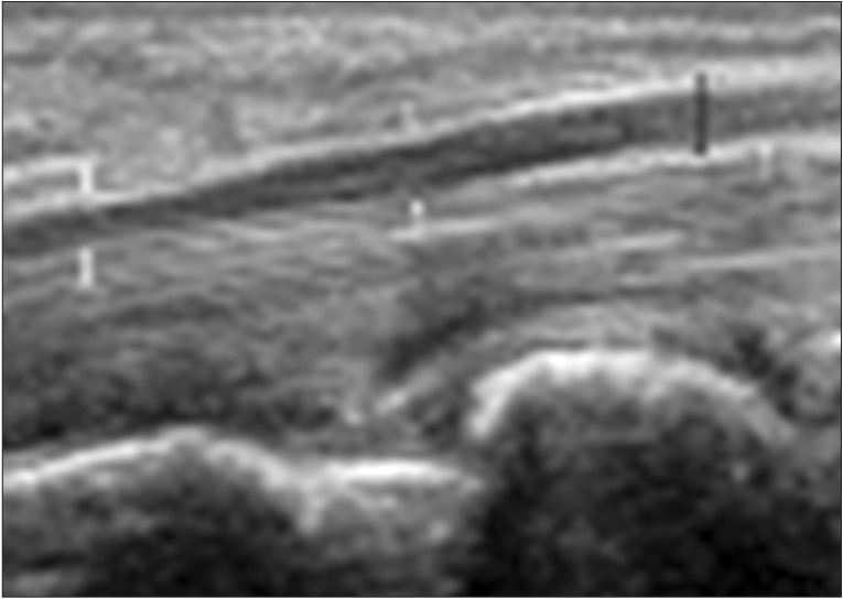
Fig. 2 Longitudinal sonogram of the median nerve (between white arrows) at the level of the carpal tunnel in a normal subject. The thickest anteroposterior diameter of the median nerve (black arrow).
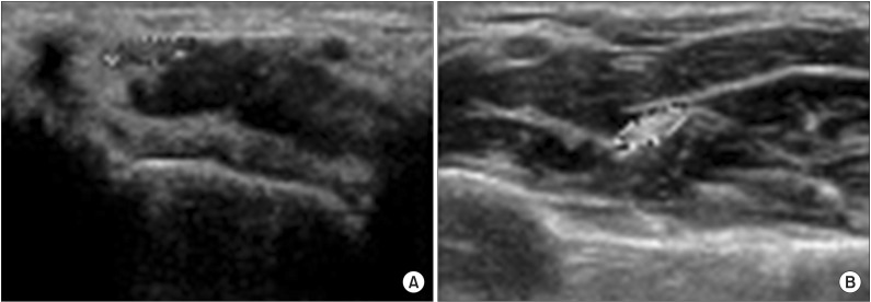
Fig. 3 Transverse sonogram of the median nerve with cross-sectional area (within oval dotted line) in a normal subject. (A) At the distal wrist crease. (B) At 12 cm proximal in the forearm. The wrist to forearm ratio was calculated as the ratio of the median nerve area at the distal wrist crease as compared to the forearm.
Reference
-
1. Phalen GS. The carpal-tunnel syndrome: clinical evaluation of 598 hands. Clin Orthop Relat Res. 1972; 83:29–40. PMID: 5014825.2. El Miedany Y, Ashour S, Youssef S, Mehanna A, Meky FA. Clinical diagnosis of carpal tunnel syndrome: old tests-new concepts. Joint Bone Spine. 2008; 75:451–457. PMID: 18455945.
Article3. Jablecki CK, Andary MT, Floeter MK, Miller RG, Quartly CA, Vennix MJ, et al. Practice parameter: Electrodiagnostic studies in carpal tunnel syndrome. Report of the American Association of Electrodiagnostic Medicine, American Academy of Neurology, and the American Academy of Physical Medicine and Rehabilitation. Neurology. 2002; 58:1589–1592. PMID: 12058083.
Article4. Buchberger W. Radiologic imaging of the carpal tunnel. Eur J Radiol. 1997; 25:112–117. PMID: 9283839.
Article5. Buchberger W, Schon G, Strasser K, Jungwirth W. High-resolution ultrasonography of the carpal tunnel. J Ultrasound Med. 1991; 10:531–537. PMID: 1942218.
Article6. Buchberger W, Judmaier W, Birbamer G, Lener M, Schmidauer C. Carpal tunnel syndrome: diagnosis with high-resolution sonography. AJR Am J Roentgenol. 1992; 159:793–798. PMID: 1529845.
Article7. Choi WK, Kang YK, Kim YH, Park EM. Diagnosis of carpal tunnel syndrome by diagnostic ultrasound. J Korean Acad Rehabil Med. 2001; 25:134–139.8. Kim MS, Yoon SY, Lee SO, Lee YG. The abnormal ultrasonographic findings of carpal tunnel in carpal tunnel syndrome. J Korean Acad Rehabil Med. 2003; 27:735–739.9. Duncan I, Sullivan P, Lomas F. Sonography in the diagnosis of carpal tunnel syndrome. AJR Am J Roentgenol. 1999; 173:681–684. PMID: 10470903.
Article10. Lee D, van Holsbeeck MT, Janevski PK, Ganos DL, Ditmars DM, Darian VB. Diagnosis of carpal tunnel syndrome: ultrasound versus electromyography. Radiol Clin North Am. 1999; 37:859–872. PMID: 10442084.11. Park GY, Bae JH, Lee SY, Oh JS, Lim JG, Son DG. Ultrasonographic findings of mild and very mild carpal tunnel syndrome. J Korean Acad Rehabil Med. 2008; 32:67–72.12. Hobson-Webb LD, Massey JM, Juel VC, Sanders DB. The ultrasonographic wrist-to-forearm median nerve area ratio in carpal tunnel syndrome. Clin Neurophysiol. 2008; 119:1353–1357. PMID: 18387336.
Article13. Wong SM, Griffith JF, Hui AC, Lo SK, Fu M, Wong KS. Carpal tunnel syndrome: diagnostic usefulness of sonography. Radiology. 2004; 232:93–99. PMID: 15155897.
Article14. Keberle M, Jenett M, Kenn W, Reiners K, Peter M, Haerten R, et al. Technical advances in ultrasound and MR imaging of carpal tunnel syndrome. Eur Radiol. 2000; 10:1043–1050. PMID: 11003395.
Article15. Kim HJ, Lee BN. Correlation of ultrasonography with the nerve conduction study in carpal tunnel syndrome of Koreans. J Korean Assoc EMG Electrodiagn Med. 2006; 8:21–25.16. Lee SJ, Kim JS, Choi YR, Kim SJ, Kang HJ. Relationship between change of median nerve cross-sectional area measured by ultrasonography and prognosis after carpal tunnel release. J Korean Orthop Assoc. 2013; 48:290–296.
Article17. Johnson EW, Melvin JL. Sensory conduction studies of median and ulnar nerves. Arch Phys Med Rehabil. 1967; 48:25–30. PMID: 6016563.18. Melvin JL, Schuchmann JA, Lanese RR. Diagnostic specificity of motor and sensory nerve conduction variables in the carpal tunnel syndrome. Arch Phys Med Rehabil. 1973; 54:69–74. PMID: 4692637.19. Johnson EW, Kukla RD, Wongsam PE, Piedmont A. Sensory latencies to the ring finger: normal values and relation to carpal tunnel syndrome. Arch Phys Med Rehabil. 1981; 62:206–208. PMID: 7235908.20. American Association of Electrodiagnostic Medicine, American Academy of Neurology, and American Academy of Physical Medicine and Rehabilitation. Practice parameter for electrodiagnostic studies in carpal tunnel syndrome: summary statement. Muscle Nerve. 2002; 25:918–922. PMID: 12115985.21. Akelman E, Weiss AP. Carpal tunnel syndrome. Etiology and endoscopic treatment. Orthop Clin North Am. 1995; 26:769–778. PMID: 7566922.22. Omer GE Jr. Median nerve compression at the wrist. Hand Clin. 1992; 8:317–324. PMID: 1613039.
Article23. Cartwright MS, Shin HW, Walker FO. Ultrasonographic characteristics of the normal median nerve. Neurology. 2006; 66(Suppl 2):A83.
- Full Text Links
-
- Actions
-
Cited
- CITED
-
- Close
- Share
-
- Similar articles
-
- Analysis of Sonographic Measurement by Anatomical Area in Carpal Tunnel Syndrome and Correlation the Measurement with Electrodiagnostic Study
- The Correlation Between Electrodiagnostic Results and Ultrasonographic Findings in the Severity of Carpal Tunnel Syndrome in Females
- Ultrasonographic Study of Median Nerve after Carpal Tunnel Release
- Carpal Tunnel Syndrome
- Diagnosis of Carpal Tunnel Syndrome by Diagnostic Ultrasound
