The Correlation Between Electrodiagnostic Results and Ultrasonographic Findings in the Severity of Carpal Tunnel Syndrome in Females
- Affiliations
-
- 1Department of Physical Medicine and Rehabilitation, National Health Insurance Service Ilsan Hospital, Goyang, Korea. rehappydoc@gmail.com
- KMID: 2389405
- DOI: http://doi.org/10.5535/arm.2017.41.4.595
Abstract
OBJECTIVE
To determine which ultrasonographic measurement can be used as an indicator reflecting the severity of carpal tunnel syndrome (CTS), by comparing electrodiagnostic results with ultrasonographic measurements in females. Many previous studies have tried to reveal that the ultrasonography (US) can possibility be used for diagnosis and severity of CTS. However, the criteria are different by gender. Thus far, there have been many efforts towards providing patients with a CTS diagnosis and severity prediction using US, but studies' results are still unclear due to lack of data on gender differences.
METHODS
We collected data from 54 female patients. We classified the severity of CTS according to electrodiagnostic results. Ultrasonographic measurements included proximal and distal cross-sectional areas of the median nerve and carpal tunnel.
RESULTS
The severity by electrodiagnostic results statistically correlated to the proximal cross-sectional area (CSA) of the median nerve and carpal tunnel. However, there was no relationship between the proximal and distal nerve/tunnel indexes and the severity by electrodiagnostic results.
CONCLUSION
In female patients with CTS, the proximal CSAs of the median nerve and carpal tunnel increase. They correlate with the severity by electrodiagnostic findings. The CSA of the proximal median nerve could be particularly used as a predictor of the severity of CTS in female patients. However, the nerve/tunnel index is constant, irrespective of the severity of CTS.
Figure
-
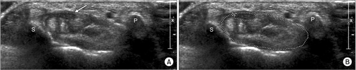
Fig. 1 Cross-sectional areas of the median nerve and carpal tunnel at proximal carpal tunnel level. (A) Proximal carpal tunnel, median nerve (arrow), and flexor tendon groups are shown between the pisiform and scaphoid bones. (B) Dotted outlines of median nerve and carpal tunnel are shown at proximal carpal tunnel level. S, means scaphoid bone; P, means pisiform bone.
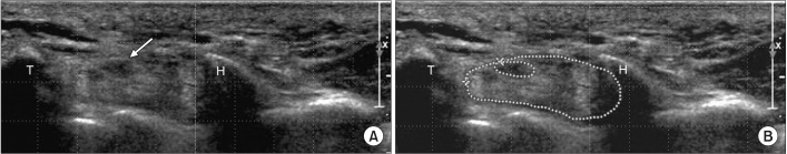
Fig. 2 Cross-sectional areas of the median nerve and carpal tunnel at distal carpal tunnel level. (A) Proximal carpal tunnel, median nerve (arrow), and flexor tendon groups are shown between the trapezium and hook of hamate bones. (B) Dotted outlines of median nerve and carpal tunnel are shown at distal carpal tunnel level. T, means trapezium bone; H, means hook of hamate bone.
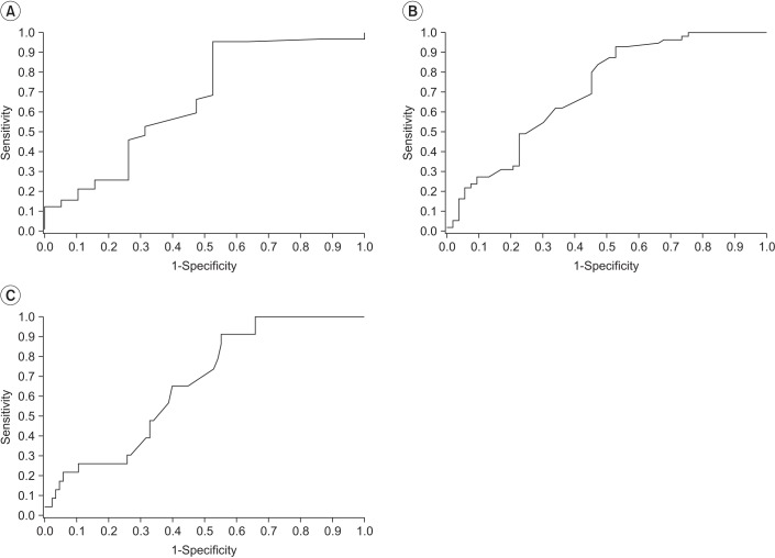
Fig. 3 Receiver operating characteristic curves of cross-sectional area of proximal median nerve. (A) Cut-off value of proximal cross-sectional area of median nerve in severity grade I is 10 mm2 (AUC=0.66, sensitivity=86.52%, 1-specificity=52.63%). (B) Cut-off value of proximal cross-sectional area of median nerve in severity grade II is 11 mm2 (AUC=0.71, sensitivity=82.27%, 1-specificity=50.94%). (C) Cut-off value of proximal cross-sectional area of median nerve in severity grade III is 12 mm2 (AUC=0.66, sensitivity=86.96%, 1-specificity=55.29%). AUC, area under the curve.
Reference
-
1. Karadag YS, Karadag O, Cicekli E, Ozturk S, Kiraz S, Ozbakir S, et al. Severity of carpal tunnel syndrome assessed with high frequency ultrasonography. Rheumatol Int. 2010; 30:761–765. PMID: 19593567.
Article2. de Krom MC, Knipschild PG, Kester AD, Thijs CT, Boekkooi PF, Spaans F. Carpal tunnel syndrome: prevalence in the general population. J Clin Epidemiol. 1992; 45:373–376. PMID: 1569433.
Article3. Duncan I, Sullivan P, Lomas F. Sonography in the diagnosis of carpal tunnel syndrome. AJR Am J Roentgenol. 1999; 173:681–684. PMID: 10470903.
Article4. Pastare D, Therimadasamy AK, Lee E, Wilder-Smith EP. Sonography versus nerve conduction studies in patients referred with a clinical diagnosis of carpal tunnel syndrome. J Clin Ultrasound. 2009; 37:389–393. PMID: 19479718.
Article5. Kim HS, Joo SH, Han ZA, Kim YW. The nerve/tunnel index: a new diagnostic standard for carpal tunnel syndrome using sonography: a pilot study. J Ultrasound Med. 2012; 31:23–29. PMID: 22215765.6. Carneiro RS, Velasquez L, Tietzman A. Trigger wrist caused by a tumor of the tendon sheath in a teenager. Am J Orthop (Belle Mead NJ). 2001; 30:233–234. PMID: 11300133.7. Bou-Merhi JS, Harris PG, Brutus JP. “Trigger finger at the wrist” due to anomalous flexor digitorum superficialis muscle belly within the carpal tunnel. Chir Main. 2007; 26:238–242. PMID: 17920326.8. Kim HS, Joo SH, Cho HK, Kim YW. Comparison of proximal and distal cross-sectional areas of the median nerve, carpal tunnel, and nerve/tunnel index in subjects with carpal tunnel syndrome. Arch Phys Med Rehabil. 2013; 94:2151–2156. PMID: 23727345.
Article9. El Miedany YM, Aty SA, Ashour S. Ultrasonography versus nerve conduction study in patients with carpal tunnel syndrome: substantive or complementary tests? Rheumatology (Oxford). 2004; 43:887–895. PMID: 15100417.
Article10. Moran L, Perez M, Esteban A, Bellon J, Arranz B, del Cerro M. Sonographic measurement of cross-sectional area of the median nerve in the diagnosis of carpal tunnel syndrome: correlation with nerve conduction studies. J Clin Ultrasound. 2009; 37:125–131. PMID: 19170107.
Article11. Claes F, Kasius KM, Meulstee J, Verhagen WI. Comparing a new ultrasound approach with electrodiagnostic studies to confirm clinically defined carpal tunnel syndrome: a prospective, blinded study. Am J Phys Med Rehabil. 2013; 92:1005–1011. PMID: 23811615.12. Mhoon JT, Juel VC, Hobson-Webb LD. Median nerve ultrasound as a screening tool in carpal tunnel syndrome: correlation of cross-sectional area measures with electrodiagnostic abnormality. Muscle Nerve. 2012; 46:871–878. PMID: 23041984.
Article13. Tai TW, Wu CY, Su FC, Chern TC, Jou IM. Ultrasonography for diagnosing carpal tunnel syndrome: a meta-analysis of diagnostic test accuracy. Ultrasound Med Biol. 2012; 38:1121–1128. PMID: 22542258.
Article14. Fowler JR, Gaughan JP, Ilyas AM. The sensitivity and specificity of ultrasound for the diagnosis of carpal tunnel syndrome: a meta-analysis. Clin Orthop Relat Res. 2011; 469:1089–1094. PMID: 20963527.
Article15. American Association of Electrodiagnostic Medicine. American Academy of Neurology. American Academy of Physical Medicine and Rehabilitation. Practice parameter for electrodiagnostic studies in carpal tunnel syndrome: summary statement. Muscle Nerve. 1993; 16:1390–1391. PMID: 8232398.16. Uncini A, Lange DJ, Solomon M, Soliven B, Meer J, Lovelace RE. Ring finger testing in carpal tunnel syndrome: a comparative study of diagnostic utility. Muscle Nerve. 1989; 12:735–741. PMID: 2641997.
Article17. Werner RA, Andary M. Electrodiagnostic evaluation of carpal tunnel syndrome. Muscle Nerve. 2011; 44:597–607. PMID: 21922474.
Article18. Bland JD. A neurophysiological grading scale for carpal tunnel syndrome. Muscle Nerve. 2000; 23:1280–1283. PMID: 10918269.
Article19. Nakamichi K, Tachibana S. Ultrasonographic measurement of median nerve cross-sectional area in idiopathic carpal tunnel syndrome: diagnostic accuracy. Muscle Nerve. 2002; 26:798–803. PMID: 12451604.
Article20. Yazdchi M, Tarzemani MK, Mikaeili H, Ayromlu H, Ebadi H. Sensitivity and specificity of median nerve ultrasonography in diagnosis of carpal tunnel syndrome. Int J Gen Med. 2012; 5:99–103. PMID: 22319247.21. Dumitru D, Amato AA, Zwarts M. Electrodiagnostic medicine. 2nd ed. Philadelphia: Hanley & Belfus;2002.22. Buchberger W, Judmaier W, Birbamer G, Lener M, Schmidauer C. Carpal tunnel syndrome: diagnosis with high-resolution sonography. AJR Am J Roentgenol. 1992; 159:793–798. PMID: 1529845.
Article23. Bianchi S, Martinoli C. Ultrasound of the musculoskeletal system. Heidelberg: Springer;2007.24. Yesildag A, Kutluhan S, Sengul N, Koyuncuoglu HR, Oyar O, Guler K, et al. The role of ultrasonographic measurements of the median nerve in the diagnosis of carpal tunnel syndrome. Clin Radiol. 2004; 59:910–915. PMID: 15451351.
Article25. Jablecki CK, Andary MT, Floeter MK, Miller RG, Quartly CA, Vennix MJ, et al. Practice parameter: electrodiagnostic studies in carpal tunnel syndrome. Report of the American Association of Electrodiagnostic Medicine, American Academy of Neurology, and the American Academy of Physical Medicine and Rehabilitation. Neurology. 2002; 58:1589–1592. PMID: 12058083.
Article
- Full Text Links
-
- Actions
-
Cited
- CITED
-
- Close
- Share
-
- Similar articles
-
- Analysis of Sonographic Measurement by Anatomical Area in Carpal Tunnel Syndrome and Correlation the Measurement with Electrodiagnostic Study
- Pressure Measurement in Carpal Tunnel Syndrome : Correlation with Electrodiagnostic and Ultrasonographic Findings
- The Relationship between Clinical and Electrodiagnostic Findings in Carpal Tunnel Syndrome
- The Correlation of Electrodiagnostic Severity, Severity of Symptom, Functional Status, and Clinical Severity in Patients with Carpal Tunnel Syndrome
- Ultrasonographic Study of Median Nerve after Carpal Tunnel Release
