Healthc Inform Res.
2017 Jan;23(1):53-59. 10.4258/hir.2017.23.1.53.
New Aging Index Using Signal Features of Both Photoplethysmograms and Acceleration Plethysmograms
- Affiliations
-
- 1Department of Electronics Engineering, College of Engineering, Hallym University, Chuncheon, Korea. ajm@hallym.ac.kr
- KMID: 2368985
- DOI: http://doi.org/10.4258/hir.2017.23.1.53
Abstract
OBJECTIVES
Acceleration plethysmograms (APGs) are obtained by taking the second derivative of photoplethysmograms (PPGs) and are noninvasive circulatory signals related to risk factors for atherosclerosis with age. There has been growing interest in the development of mobile devices to collect and analyze PPG single features for ambulatory health monitoring. The present study aimed to extract a new feature from the morphologies of APG and PPG signals to classify the dominant indices related to the pulsatile volume of blood in tissue according to age.
METHODS
Ten APG and 14 PPG indices were simultaneously extracted. All indices were compared via Pearson correlation coefficients (r) and a regression analysis. We introduced a combined index extracted from both the PPG and APG indices defined as the inflection point area plus the d_peak (IPAD). The participants included 93 healthy adults aged 36-86 years with a mean ± standard deviation age of 57.43 ± 11.99 years.
RESULTS
The d_peak and age index for the APG indices were significantly correlated with age (r = −0.408, p < 0.0001 and r = 0.296, p = 0.0039, respectively). Only the A1 time for PPG indices was moderately correlated with age (r = −0.247, p = 0.017). The stiffness index, including individual height information, was not related to age (r = −0.031, p = 0.7713). However, the combined IPAD index was significantly more correlated with age (r = 0.56, p < 0.001) than the other indices.
CONCLUSIONS
The proposed index outperformed the other 24 indices for evaluating vascular aging. We suggest that the IPAD is a significant factor related to the clinical information embedded in the PPG waveform.
Keyword
MeSH Terms
Figure
-
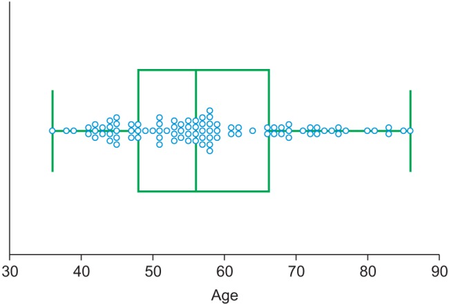
Figure 1 Age group distribution.
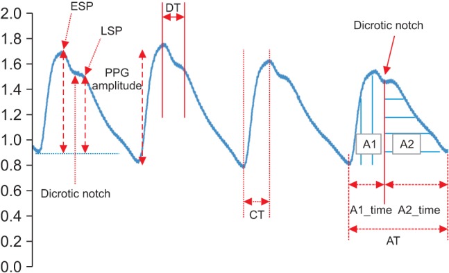
Figure 2 Photoplethysmogram (PPG) signal features. ESP: early systolic peak, LSP: late systolic peak, DT: delta time, CT: crest time, AT: area time.
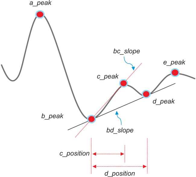
Figure 3 Acceleration plethysmogram signal features.
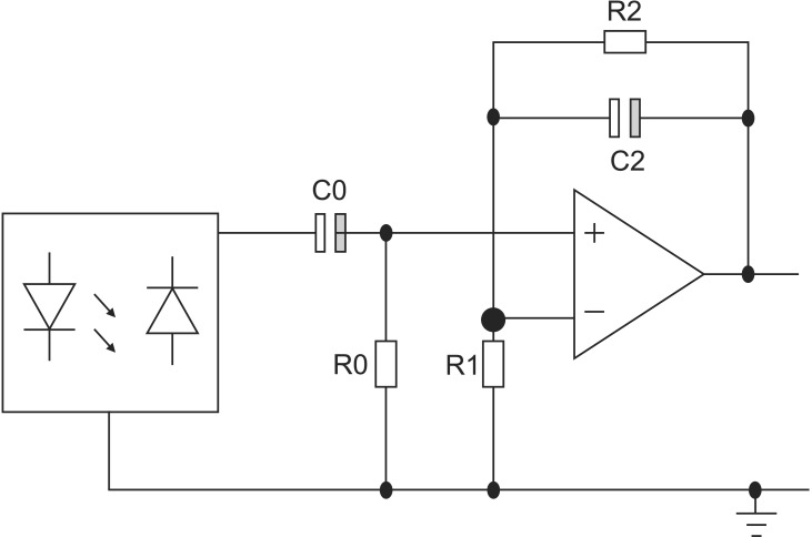
Figure 4 Circuit topology of the preamplifier.
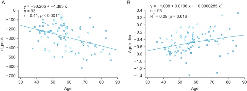
Figure 5 (A) The relationship between the peak and age and (B) the relationship between the age index and age.
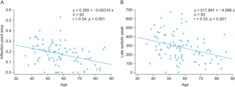
Figure 6 (A) The relationship between age and inflection point area (IPA) and (B) the relationship between age and the age index.
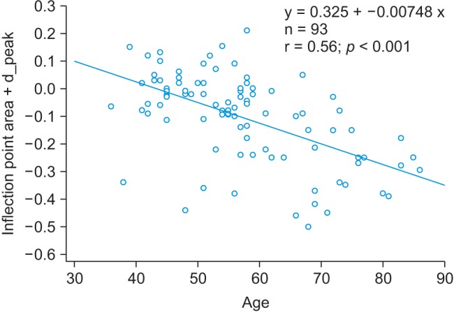
Figure 7 The relationship between age and the inflection point area plus the d_peak (IPAD).
Reference
-
1. Krishnan R, Natarajan BB, Warren S. Two-stage approach for detection and reduction of motion artifacts in photoplethysmographic data. IEEE Trans Biomed Eng. 2010; 57(8):1867–1876. PMID: 20172800.
Article2. Hamner JW, Tan CO, Lee K, Cohen MA, Taylor JA. Sympathetic control of the cerebral vasculature in humans. Stroke. 2010; 41(1):102–109. PMID: 20007920.
Article3. Chellappan K, Ali MM, Zahedi E. An age index for vascular system based on photoplethysmogram pulse contour analysis. Proceedings of 4th Kuala Lumpur International Conference on Biomedical Engineering. 2008 Jun 25-28; Kuala Lumpur, Malaysia. p. 125–128.4. Yousef Q, Reaz MB, Ali MA. The analysis of PPG morphology: investigating the effects of aging on arterial compliance. Meas Sci Rev. 2012; 12(6):266–271.
Article5. Elgendi M. On the analysis of fingertip photoplethysmogram signals. Curr Cardiol Rev. 2012; 8(1):14–25. PMID: 22845812.
Article6. Suganthi L, Manivannan M, Kunwar BK, Joseph G, Danda D. Morphological analysis of peripheral arterial signals in Takayasu's arteritis. J Clin Monit Comput. 2015; 29(1):87–95. PMID: 24652647.
Article7. Ahn JM. Wave detection in acceleration plethysmogram. Healthc Inform Res. 2015; 21(2):111–117. PMID: 25995963.
Article8. Dorlas JC, Nijboer JA. Photo-electric plethysmography as a monitoring device in anaesthesia: application and interpretation. Br J Anaesth. 1985; 57(5):524–530. PMID: 3994887.9. Millasseau SC, Kelly RP, Ritter JM, Chowienczyk PJ. Determination of age-related increases in large artery stiffness by digital pulse contour analysis. Clin Sci (Lond). 2002; 103(4):371–377. PMID: 12241535.
Article10. Padilla JM, Berjano EJ, Saiz J, Facila L, Diaz P, Merce S. Assessment of relationships between blood pressure, pulse wave velocity and digital volume pulse. Proceedings of 2006 33rd Conference on Computers in Cardiology. 2006 Sep 17-20; Valencia, Spain. p. 893–896.11. Rubins U, Grabovskis A, Grube J, Kukulis I. Photoplethysmography analysis of artery properties in patients with cardiovascular diseases. Proceedings of 14th Nordic-Baltic Conference on Biomedical Engineering and Medical Physics. 2008 Jun 16-20; Riga, Latvia. p. 319–322.12. Wang L, Pickwell-MacPherson E, Liang YP, Zhang YT. Noninvasive cardiac output estimation using a novel photoplethysmogram index. Proceedings of 31st Annual International Conference of the IEEE Engineering in Medicine and Biology Society. 2009 Sep 2-6; Minneapolis, MN. p. 1746–1749.13. Takazawa K, Fujita M, Yabe K, Sasaki T, Kobayashi T, Maeda K. Clinical usefulness of the second derivative of a plethysmogram. J Cardiol. 1993; 23:207–217.14. Takazawa K, Tanaka N, Fujita M, Matsuoka O, Saiki T, Aikawa M, et al. Assessment of vasoactive agents and vascular aging by the second derivative of photoplethysmogram waveform. Hypertension. 1998; 32(2):365–370. PMID: 9719069.
Article15. Inuma J, Murakoshi M, Kobayashi T, Io H, Kaneko K, Takahashi T, et al. Relationship between acceleration plethysmography and aortic calcification index in chronic kidney disease patients. Hong Kong J Nephrol. 2012; 14(2):48–53.
Article16. Hong KS, Park KT, Ahn JM. Aging index using photoplethysmography for a healthcare device: comparison with brachial-ankle pulse wave velocity. Healthc Inform Res. 2015; 21(1):30–34. PMID: 25705555.
Article17. Tomiyama H, Yamashina A, Arai T, Hirose K, Koji Y, Chikamori T, et al. Influences of age and gender on results of noninvasive brachial-ankle pulse wave velocity measurement: a survey of 12517 subjects. Atherosclerosis. 2003; 166(2):303–309. PMID: 12535743.18. Munakata M, Nunokawa T, Yoshinaga K, Toyota T. The brachial-ankle pulse wave velocity is a better predictor for pulse pressure than augmentation index in older hypertensives. Japan Med Assoc J. 2005; 48(5):224–233.
- Full Text Links
-
- Actions
-
Cited
- CITED
-
- Close
- Share
-
- Similar articles
-
- Wave Detection in Acceleration Plethysmogram
- Aging Index using Photoplethysmography for a Healthcare Device: Comparison with Brachial-Ankle Pulse Wave Velocity
- The Effect of the Signal Loss on the Fetal Heart Rate Variables
- Changes of Left Ventricular Diastolic Function in Normal Children
- Growth Hormone and Aging: Updated Review
