Comparison of the fracture resistances of glass fiber mesh- and metal mesh-reinforced maxillary complete denture under dynamic fatigue loading
- Affiliations
-
- 1Department of Prosthodontics and Research Institute of Oral Science, College of Dentistry, Gangneung-Wonju National University, Gangneung, Republic of Korea. doctorcj@gwnu.ac.kr
- KMID: 2368208
- DOI: http://doi.org/10.4047/jap.2017.9.1.22
Abstract
- PURPOSE
The aim of this study was to investigate the effect of reinforcing materials on the fracture resistances of glass fiber mesh- and Cr-Co metal mesh-reinforced maxillary complete dentures under fatigue loading.
MATERIALS AND METHODS
Glass fiber mesh- and Cr-Co mesh-reinforced maxillary complete dentures were fabricated using silicone molds and acrylic resin. A control group was prepared with no reinforcement (n = 15 per group). After fatigue loading was applied using a chewing simulator, fracture resistance was measured by a universal testing machine. The fracture patterns were analyzed and the fractured surfaces were observed by scanning electron microscopy.
RESULTS
After cyclic loading, none of the dentures showed cracks or fractures. During fracture resistance testing, all unreinforced dentures experienced complete fracture. The mesh-reinforced dentures primarily showed posterior framework fracture. Deformation of the all-metal framework caused the metal mesh-reinforced denture to exhibit the highest fracture resistance, followed by the glass fiber mesh-reinforced denture (P<.05) and the control group (P<.05). The glass fiber mesh-reinforced denture primarily maintained its original shape with unbroken fibers. River line pattern of the control group, dimples and interdendritic fractures of the metal mesh group, and radial fracture lines of the glass fiber group were observed on the fractured surfaces.
CONCLUSION
The glass fiber mesh-reinforced denture exhibits a fracture resistance higher than that of the unreinforced denture, but lower than that of the metal mesh-reinforced denture because of the deformation of the metal mesh. The glass fiber mesh-reinforced denture maintains its shape even after fracture, indicating the possibility of easier repair.
MeSH Terms
Figure
-
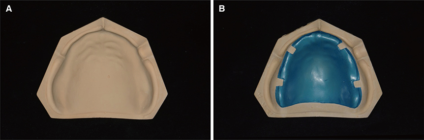
Fig. 1 (A) Maxillary edentulous master cast, (B) 0.5 mm relief on cast.
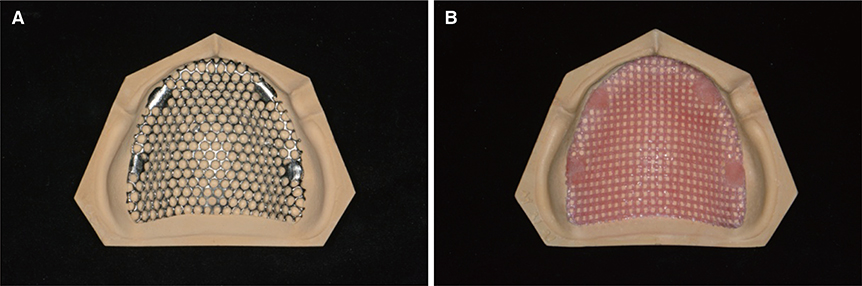
Fig. 2 (A) Metal mesh framework, (B) Glass fiber mesh framework on cast.
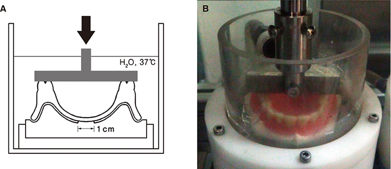
Fig. 3 (A) Schematic representation of loading for experimental design. The arrow indicates the direction of the cyclic force, (B) Chewing simulator.
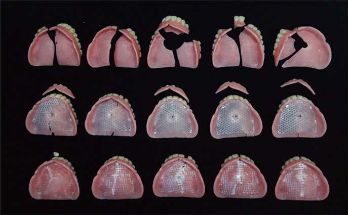
Fig. 4 Representative fracture patterns of complete dentures. Upper: resin complete dentures, Middle: metal meshreinforced complete dentures, Lower: fiber mesh-reinforced complete dentures.
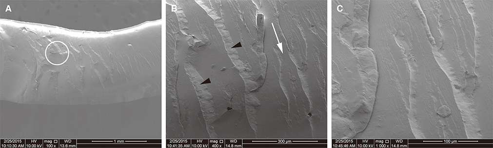
Fig. 5 Fractured surface of the unreinforced resin complete denture; palate area of acrylic resin base. (A) White circle indicates pink fibers (× 100), (B), (C) River line pattern in higher magnification (× 400 and × 1,000). White arrow indicates direction of the river line propagation. Black points indicate scarps.
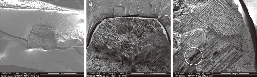
Fig. 6 Fractured surface of the metal mesh-reinforced complete denture; palate area of acrylic resin base. (A) Horizontal crack line (× 100). Trapezoidal material of the center is a part of metal mesh. (B) Gap between metal and acrylic resin. And river line pattern of acrylic resin (× 400). (C) Dimple and interdendritic fracture. White circle represents shrinkage cavity formation in casting (× 1,000).
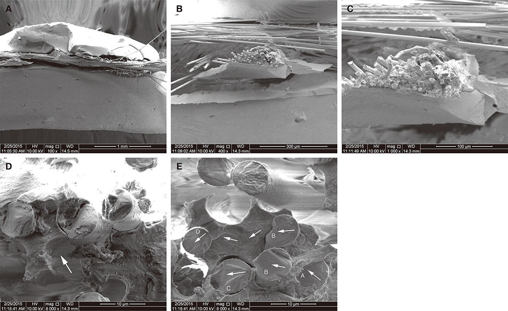
Fig. 7 Fractured surface of the glass fiber mesh-reinforced complete denture; palate area of acrylic resin base. (A) Crisscross fibers are broken (× 100), (B) Broken acrylic resin and broom-like fiber ends (× 400), (C) Broken glass fibers (× 1,000), (D) Cracked fiber ends in resin matrix. White arrow represents clean separation of the fiber from the matrix (× 8,000), (E) The fiber ends exhibit radial features and acrylic resin shows the microflow. From this, white arrow represents direction of fractures (× 8,000).
Reference
-
1. Jagger DC, Harrison A, Jandt KD. The reinforcement of dentures. J Oral Rehabil. 1999; 26:185–194.2. el Ghazali S, Glantz PO, Strandman E, Randow K. On the clinical deformation of maxillary complete dentures. Influence of denture-base design and shape of denture-bearing tissue. Acta Odontol Scand. 1989; 47:69–76.3. Geurtsen W. Biocompatibility of dental casting alloys. Crit Rev Oral Biol Med. 2002; 13:71–84.4. Kim SH, Watts DC. The effect of reinforcement with woven E-glass fibers on the impact strength of complete dentures fabricated with high-impact acrylic resin. J Prosthet Dent. 2004; 91:274–280.5. Hedzelek W, Gajdus P. Mechanical strength of an acrylic resin palatal denture base reinforced with a mesh or bundle of glass fibers. Int J Prosthodont. 2007; 20:311–312.6. Yu SH, Cho HW, Oh S, Bae JM. Effects of glass fiber mesh with different fiber content and structures on the compressive properties of complete dentures. J Prosthet Dent. 2015; 113:636–644.7. Takahashi Y, Yoshida K, Shimizu H. Effect of location of glass fiber-reinforced composite reinforcement on the flexural properties of a maxillary complete denture in vitro. Acta Odontol Scand. 2011; 69:215–221.8. Vallittu PK. Flexural properties of acrylic resin polymers reinforced with unidirectional and woven glass fibers. J Prosthet Dent. 1999; 81:318–326.9. Vallittu PK, Lassila VP, Lappalainen R. Transverse strength and fatigue of denture acrylic-glass fiber composite. Dent Mater. 1994; 10:116–121.10. Vallittu PK. A review of fiber-reinforced denture base resins. J Prosthodont. 1996; 5:270–276.11. Fontijn-Tekamp FA, Slagter AP, Van Der Bilt A, Van ‘T Hof MA, Witter DJ, Kalk W, Jansen JA. Biting and chewing in overdentures, full dentures, and natural dentitions. J Dent Res. 2000; 79:1519–1524.12. Michael CG, Javid NS, Colaizzi FA, Gibbs CH. Biting strength and chewing forces in complete denture wearers. J Prosthet Dent. 1990; 63:549–553.13. Bates JF, Stafford GD, Harrison A. Masticatory function-a review of the literature: (II) Speed of movement of the mandible, rate of chewing and forces developed in chewing. J Oral Rehabil. 1975; 2:349–361.14. Wiskott HW, Nicholls JI, Belser UC. Stress fatigue: basic principles and prosthodontic implications. Int J Prosthodont. 1995; 8:105–116.15. Praveen B, Babaji HV, Prasanna BG, Rajalbandi SK, Shreeharsha TV, Prashant GM. Comparison of impact strength and fracture morphology of different heat cure denture acrylic resins: An in vitro study. J Int Oral Health. 2014; 6:12–16.16. Purslow D. Matrix fractography of fibre-reinforced epoxy composites. Composites. 1986; 17:289–303.17. Dharmar S, Rathnasamy RJ, Swaminathan TN. Radiographic and metallographic evaluation of porosity defects and grain structure of cast chromium cobalt removable partial dentures. J Prosthet Dent. 1993; 69:369–373.18. Kuroda S, Yokoyama D, Shinya A, Gomi H, Shinya A. Measuring the effects of water immersion conditions on the durability of fiber-reinforced hybrid composite resin using static and dynamic tests. Dent Mater J. 2012; 31:449–457.
- Full Text Links
-
- Actions
-
Cited
- CITED
-
- Close
- Share
-
- Similar articles
-
- Comparison of metal wire reinforcement and glass fiber reinforcement in repaired maxillary complete denture
- Effect of location of glass fiber pre-impregnated with light-curing resin on the fracture strength and fracture modes of a maxillary complete denture
- Comparison of Titanium Micro Mesh(R) with Titanium Mesh Screen 1.3(R) in the Reconstruction of Medial Orbital wall Fracture
- Evaluation of static fracture resistances and patterns of pulpless tooth restored with poly-ether-ketone-ketone (PEKK) post
- Comparison of Porous Polyethylene Sheet(Medpor(R)) with Titanium Dynamic Mesh in the Treatment of Blow Out Fracture
