Retreatability of two endodontic sealers, EndoSequence BC Sealer and AH Plus: a micro-computed tomographic comparison
- Affiliations
-
- 1Department of Endodontics, University of Washington, Seattle, WA, USA. avina@uw.edu
- 2Department of Pediatrics (Craniofacial Medicine), University of Washington, Seattle, WA, USA.
- 3Center for Developmental Biology and Regenerative Medicine, Seattle Children's Research Institute, Seattle, WA, USA.
- 4Department of Anatomy and Developmental Biology, Monash University, Clayton, Victoria, Australia.
- KMID: 2367317
- DOI: http://doi.org/10.5395/rde.2017.42.1.19
Abstract
OBJECTIVES
Recently, bioceramic sealers like EndoSequence BC Sealer (BC Sealer) have been introduced and are being used in endodontic practice. However, this sealer has limited research related to its retreatability. Hence, the aim of this study was to evaluate the retreatability of two sealers, BC Sealer as compared with AH Plus using micro-computed tomographic (micro-CT) analysis.
MATERIALS AND METHODS
Fifty-six extracted human maxillary incisors were instrumented and randomly divided into 4 groups of 14 teeth: 1A, gutta-percha, AH Plus retreated with chloroform; 1B, gutta-percha, AH Plus retreated without chloroform; 2A, gutta-percha, EndoSequence BC Sealer retreated with chloroform; 2B, gutta-percha, EndoSequence BC Sealer retreated without chloroform. Micro-CT scans were taken before and after obturation and retreatment and analyzed for the volume of residual material. The specimens were longitudinally sectioned and digitized images were taken with the dental operating microscope. Data was analyzed using an ANOVA and a post-hoc Tukey test. Fisher exact tests were performed to analyze the ability to regain patency.
RESULTS
There was significantly less residual root canal filling material in the AH Plus groups retreated with chloroform as compared to the others. The BC Sealer samples retreated with chloroform had better results than those retreated without chloroform. Furthermore, patency could be re-established in only 14% of teeth in the BC Sealer without chloroform group.
CONCLUSION
The results of this study demonstrate that the BC Sealer group had significantly more residual filling material than the AH Plus group regardless of whether or not both sealers were retreated with chloroform.
Keyword
MeSH Terms
Figure
-
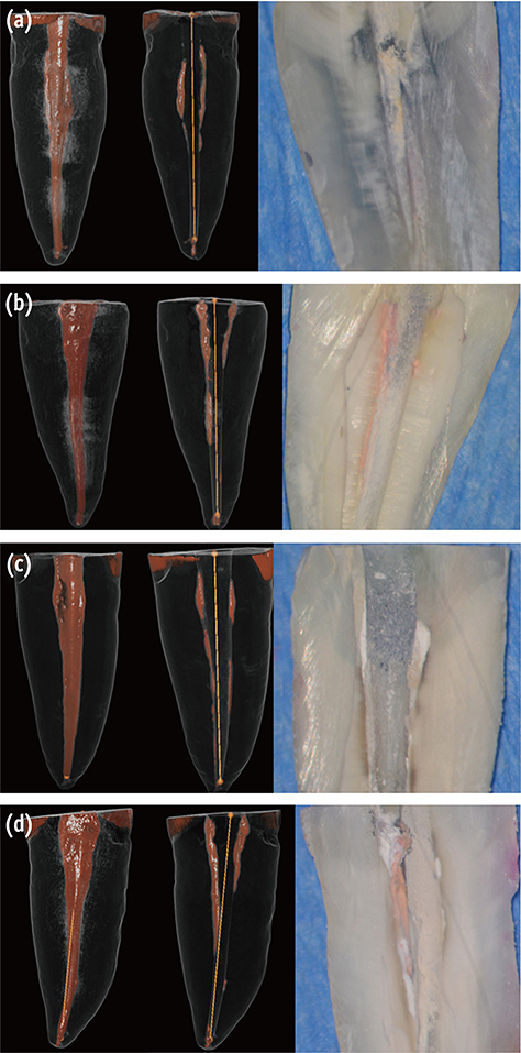
Figure 1 Visualization of residual sealer after retreatment. Micro-CT scans of obturated canals with (a) gutta-percha and AH Plus retreated with chloroform; (b) gutta-percha and AH Plus retreated without chloroform; (c) gutta-percha and BC Sealer retreated with chloroform; (d) gutta-percha and BC Sealer retreated without chloroform retreatment. The left-most images are representative 3D renderings of the filled canals, while the adjacent images are the same teeth following retreatment. Black, root surface; white and orange, obturation material. The right-most images are the corresponding longitudinal sections of these teeth. Micro-CT, micro-computed tomography; BC Sealer, EndoSequence BC Sealer (Brasseler USA, Savannah, GA, USA); 3D, 3 dimensional.
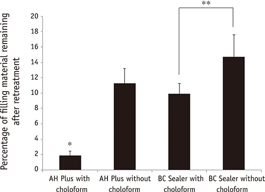
Figure 2 Percentage of residual filling material in the entire length of root canal. The differences in the percentage of residual root canal filling were statistically significant between the AH Plus with chloroform and the other groups (*p ≤ 0.05). The BC Sealer with chloroform group was significantly different from the BC Sealer without chloroform group (**p ≤ 0.05). BC Sealer, EndoSequence BC Sealer (Brasseler USA, Savannah, GA, USA).
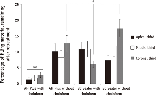
Figure 3 Percentage of residual filling material of apical third compared with middle and coronal third of canals. The differences in the percentage of residual root canal filling were statistically significant between the AH Plus with chloroform and the other groups (**p ≤ 0.05) and between the BC Sealer group retreated with chloroform and the AH Plus and BC Sealer groups that were not retreated with chloroform (*p ≤ 0.05). BC Sealer, EndoSequence BC Sealer (Brasseler USA, Savannah, GA, USA).
Cited by 5 articles
-
Micro-computed tomographic evaluation of the flow and filling ability of endodontic materials using different test models
Fernanda Ferrari Esteves Torres, Juliane Maria Guerreiro-Tanomaru, Gisselle Moraima Chavez-Andrade, Jader Camilo Pinto, Fábio Luiz Camargo Villela Berbert, Mario Tanomaru-Filho
Restor Dent Endod. 2020;45(2):e11. doi: 10.5395/rde.2020.45.e11.Effect of ultrasonic cleaning on the bond strength of fiber posts in oval canals filled with a premixed bioceramic root canal sealer
Fernando Peña Bengoa, Maria Consuelo Magasich Arze, Cristobal Macchiavello Noguera, Luiz Felipe Nunes Moreira, Augusto Shoji Kato, Carlos Eduardo Da Silveira Bueno
Restor Dent Endod. 2020;45(2):e19. doi: 10.5395/rde.2020.45.e19.Micro-computed tomographic evaluation of a new system for root canal filling using calcium silicate-based root canal sealers
Mario Tanomaru-Filho, Fernanda Ferrari Esteves Torres, Jader Camilo Pinto, Airton Oliveira Santos-Junior, Karina Ines Medina Carita Tavares, Juliane Maria Guerreiro-Tanomaru
Restor Dent Endod. 2020;45(3):e34. doi: 10.5395/rde.2020.45.e34.A micro-computed tomographic study of remaining filling materials of two bioceramic sealers and epoxy resin sealer after retreatment
KyungJae Kim, Da Vin Kim, Sin-Young Kim, SungEun Yang
Restor Dent Endod. 2019;44(2):. doi: 10.5395/rde.2019.44.e18.Efficacy of retreatment NiTi files for root canals filled with calcium silicate-based sealer
Jae-Yun Hyun, Kyung-Mo Cho, Se-Hee Park, Yoon Lee, Yoon-Joo Lee, Jin-Woo Kim
J Dent Rehabil Appl Sci. 2022;38(4):213-221. doi: 10.14368/jdras.2022.38.4.213.
Reference
-
1. Haapasalo M, Shen Y, Qian W, Gao Y. Irrigation in endodontics. Dent Clin North Am. 2010; 54:291–312.
Article2. Ng YL, Mann V, Rahbaran S, Lewsey J, Gulabivala K. Outcome of primary root canal treatment: systematic review of the literature - part 2. influence of clinical factors. Int Endod J. 2008; 41:6–31.
Article3. Ng YL, Mann V, Rahbaran S, Lewsey J, Gulabivala K. Outcome of primary root canal treatment: systematic review of the literature - part 1. effects of study characteristics on probability of success. Int Endod J. 2007; 40:921–939.
Article4. Friedman S. Prognosis of initial endodontic therapy. Endod Topics. 2002; 2:59–88.
Article5. Stabholz A, Friedman S. Endodontic retreatment-case selection and technique. Part 2: treatment planning for retreatment. J Endod. 1988; 14:607–614.
Article6. Yadav P, Bharath MJ, Sahadev CK, Makonahalli Ramachandra PK, Rao Y, Ali A, Mohamed S. An in vitro CT comparison of gutta-percha removal with two rotary systems and hedstrom files. Iran Endod J. 2013; 8:59–64.7. Barletta FB, Rahde Nde M, Limongi O, Moura AA, Zanesco C, Mazocatto G. In vitro comparative analysis of 2 mechanical techniques for removing gutta-percha during retreatment. J Can Dent Assoc. 2007; 73:65.8. Hammad M, Qualtrough A, Silikas N. Three-dimensional evaluation of effectiveness of hand and rotary instrumentation for retreatment of canals filled with different materials. J Endod. 2008; 34:1370–1373.
Article9. Kratchman SI. Obturation of the root canal system. Dent Clin North Am. 2004; 48:203–215.
Article10. Flores DS, Rached FJ Jr, Versiani MA, Guedes DF, Sousa-Neto MD, Pécora JD. Evaluation of physicochemical properties of four root canal sealers. Int Endod J. 2011; 44:126–135.
Article11. Marin-Bauza GA, Silva-Sousa YT, da Cunha SA, Rached-Junior FJ, Bonetti-Filho I, Sousa-Neto MD, Miranda CE. Physicochemical properties of endodontic sealers of different bases. J Appl Oral Sci. 2012; 20:455–461.
Article12. Hess D, Solomon E, Spears R, He J. Retreatability of a bioceramic root canal sealing material. J Endod. 2011; 37:1547–1549.
Article13. Ozkocak I, Sonat B. Evaluation of effects on the adhesion of various root canal sealers after Er:YAG laser and irrigants are used on the dentin surface. J Endod. 2015; 41:1331–1336.
Article14. Pawar SS, Pujar MA, Makandar SD. Evaluation of the apical sealing ability of bioceramic sealer, AH plus & epiphany: an in vitro study. J Conserv Dent. 2014; 17:579–582.
Article15. Topçuoğlu HS, Tuncay Ö, Karataş E, Arslan H, Yeter K. In vitro fracture resistance of roots obturated with epoxy resin-based, mineral trioxide aggregate-based, and bioceramic root canal sealers. J Endod. 2013; 39:1630–1633.
Article16. Loushine BA, Bryan TE, Looney SW, Gillen BM, Loushine RJ, Weller RN, Pashley DH, Tay FR. Setting properties and cytotoxicity evaluation of a premixed bioceramic root canal sealer. J Endod. 2011; 37:673–677.
Article17. Candeiro GT, Moura-Netto C, D'Almeida-Couto RS, Azambuja-Júnior N, Marques MM, Cai S, Gavini G. Cytotoxicity, genotoxicity and antibacterial effectiveness of a bioceramic endodontic sealer. Int Endod J. 2015; 08. 17. DOI: 10.1111/iej.12523. [Epub ahead of print].
Article18. Uzunoglu E, Yilmaz Z, Sungur DD, Altundasar E. Retreatability of root canals obturated using gutta-percha with bioceramic, MTA and resin-based sealers. Iran Endod J. 2015; 10:93–98.19. Du T, Wang Z, Shen Y, Ma J, Cao Y, Haapasalo M. Combined antibacterial effect of sodium hypochlorite and root canal sealers against Enterococcus faecalis biofilms in dentin canals. J Endod. 2015; 41:1294–1298.
Article20. Candeiro GT, Correia FC, Duarte MA, Ribeiro-Siqueira DC, Gavini G. Evaluation of radiopacity, pH, release of calcium ions, and flow of a bioceramic root canal sealer. J Endod. 2012; 38:842–845.
Article21. Gade VJ, Belsare LD, Patil S, Bhede R, Gade JR. Evaluation of push-out bond strength of endosequence BC sealer with lateral condensation and thermoplasticized technique: an in vitro study. J Conserv Dent. 2015; 18:124–127.
Article22. Wolcott JF, Himel VT, Hicks ML. Thermafil retreatment using a new ‘System B’ technique or a solvent. J Endod. 1999; 25:761–764.
Article23. Zmener O, Pameijer CH, Banegas G. Retreatment efficacy of hand versus automated instrumentation in oval-shaped root canals: an ex vivo study. Int Endod J. 2006; 39:521–526.
Article24. Ng YL, Mann V, Gulabivala K. A prospective study of the factors affecting outcomes of nonsurgical root canal treatment: part 1: periapical health. Int Endod J. 2011; 44:583–609.
Article25. Stabholz A, Friedman S. Endodontic retreatment-case selection and technique. Part 2: treatment planning for retreatment. J Endod. 1988; 14:607–614.
Article26. Friedman S, Stabholz A. Endodontic retreatment-case selection and technique. Part 1: criteria for case selection. J Endod. 1986; 12:28–33.
Article27. Rechenberg DK, Paqué F. Impact of cross-sectional root canal shape on filled canal volume and remaining root filling material after retreatment. Int Endod J. 2013; 46:547–555.
Article28. Schäfer E, Zandbiglari T. A comparison of the effectiveness of chloroform and eucalyptus oil in dissolving root canal sealers. Oral Surg Oral Med Oral Pathol Oral Radiol Endod. 2002; 93:611–616.
Article29. Bodrumlu E, Er O, Kayaoglu G. Solubility of root canal sealers with different organic solvents. Oral Surg Oral Med Oral Pathol Oral Radiol Endod. 2008; 106:e67–e69.
Article30. Han L, Okiji T. Bioactivity evaluation of three calcium silicate-based endodontic materials. Int Endod J. 2013; 46:808–814.
Article31. Han L, Okiji T. Uptake of calcium and silicon released from calcium silicate-based endodontic materials into root canal dentine. Int Endod J. 2011; 44:1081–1087.
Article32. Ersev H, Yilmaz B, Dinçol ME, Dağlaroğlu R. The efficacy of ProTaper Universal rotary retreatment instrumentation to remove single gutta-percha cones cemented with several endodontic sealers. Int Endod J. 2012; 45:756–762.
Article33. Roggendorf MJ, Legner M, Ebert J, Fillery E, Frankenberger R, Friedman S. Micro-CT evaluation of residual material in canals filled with Activ GP or GuttaFlow following removal with NiTi instruments. Int Endod J. 2010; 43:200–209.
Article34. Sağlam BC, Koçak MM, Türker SA, Koçak S. Efficacy of different solvents in removing gutta-percha from curved root canals: a micro-computed tomography study. Aust Endod J. 2014; 40:76–80.
Article35. Davidson IW, Sumner DD, Parker JC. Chloroform: a review of its metabolism, teratogenic, mutagenic, and carcinogenic potential. Drug Chem Toxicol. 1982; 5:1–87.
Article36. Margelos J, Verdelis K, Eliades G. Chloroform uptake by gutta-percha and assessment of its concentration in air during the chloroform-dip technique. J Endod. 1996; 22:547–550.
Article37. Chutich MJ, Kaminski EJ, Miller DA, Lautenschlager EP. Risk assessment of the toxicity of solvents of gutta-percha used in endodontic retreatment. J Endod. 1998; 24:213–216.
Article38. McDonald MN, Vire DE. Chloroform in the endodontic operatory. J Endod. 1992; 18:301–303.
Article39. Edgar SW, Marshall JG, Baumgartner JC. The antimicrobial effect of chloroform on Enterococcus faecalis after gutta-percha removal. J Endod. 2006; 32:1185–1187.
Article40. Tamse A, Unger U, Metzger Z, Rosenberg M. Gutta-percha solvents-a comparative study. J Endod. 1986; 12:337–339.
Article41. Hess D, Solomon E, Spears R, He J. Retreatability of a bioceramic root canal sealing material. J Endod. 2011; 37:1547–1549.
Article42. Koch KA, Brave D. EndoSequence: melding endodontics with restorative dentistry, part 3. Dent Today. 2009; 28:88–92.43. Çanakçi BC, Er O, Dincer A. Do the sealer solvents used affect apically extruded debris in retreatment? J Endod. 2015; 41:1507–1509.
Article44. Bayram E, Dalat D, Bayram M. Solubility evaluation of different root canal sealing materials. J Contemp Dent Pract. 2015; 16:96–100.
Article
- Full Text Links
-
- Actions
-
Cited
- CITED
-
- Close
- Share
-
- Similar articles
-
- Micro-computed tomographic evaluation of single-cone obturation with three sealers
- A micro-computed tomographic study of remaining filling materials of two bioceramic sealers and epoxy resin sealer after retreatment
- Cytocompatibility and cell proliferation evaluation of calcium phosphate-based root canal sealers
- Physical properties of a new resin-based root canal sealer in comparison with AH Plus Jet
- A scientometric, bibliometric, and thematic map analysis of hydraulic calcium silicate root canal sealers
