Perinatology.
2016 Dec;27(4):227-235. 10.14734/PN.2016.27.4.227.
Dose Brain MRI before Discharge at NICU Predict Neurodevelopmental Outcomes in Very Low Birth Weight Infants?
- Affiliations
-
- 1Department of Pediatrics, Seoul National University Hospital, Seoul, Korea.
- 2Department of Pediatrics, Seoul National University Bundang Hospital, Seongnam, Korea. choicw1029@gmail.com
- 3Department of Radiology, Seoul National University Bundang Hospital, Seongnam, Korea.
- KMID: 2367034
- DOI: http://doi.org/10.14734/PN.2016.27.4.227
Abstract
- PURPOSE
To test whether brain MRI can predict neurodevelopmental outcomes of very low birth weight (VLBW) infants in a single academic center.
METHODS
This was a retrospective study of VLBW infants admitted to neonatal intensive care unit from January 2010 to December 2014. Infants who were taken brain MRI before discharge and followed-up at 12 or 24 months' corrected age (CA) were enrolled. The neurodevelopment outcomes included cerebral palsy (CP) and cognitive or motor delay on Bayley Scale of Infant Development-II.
RESULTS
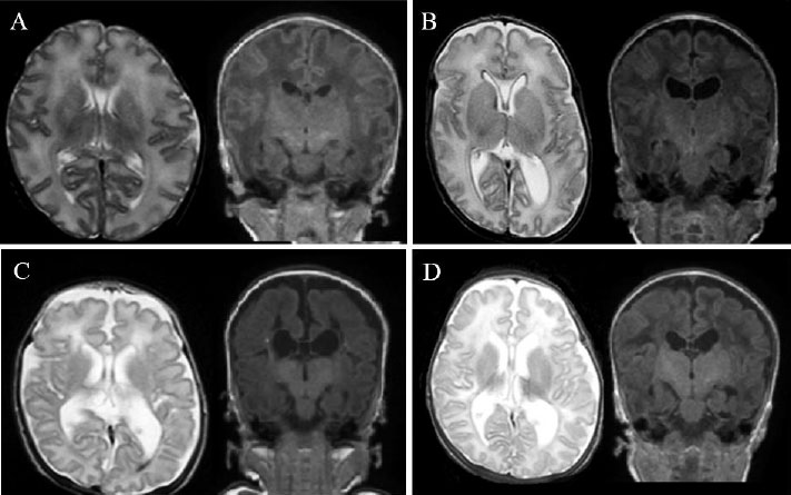
Of the 255 survivors at discharge, 182 (71.4%) had a brain MRI. Any abnormalities on brain MRI were predictive of CP (odds ratio [OR] 15.8, 95% confidence interval [CI] 1.9-128.1) and motor delay(OR 4.4, 95% CI 1.0-19.3) at 12 months' CA. Moderate to severe white matter abnormalities on brain MRI were significantly correlated with CP (OR 49.0, 95% CI 10.1-238.2) and moderate to severe motor delay (OR 8.3, 95% CI 1.2-56.7) at 12 months' CA, and CP (OR 43.8, 95% CI 6.4-299.8) at 24 months' CA. Moderate to severe white matter abnormalities on brain MRI were consistently associated with CP at 12 and 24 months' CA after adjustment for demographic and clinical variables and cranial ultrasonography findings (OR 800.5, 95% CI 6.9-92,665.7 at 12 months' CA, OR 52.0, 95% CI 1.3-2,168.2 at 24 months' CA).
CONCLUSION
Moderate to severe white matter abnormalities on brain MRI strongly predicted cerebral palsy at 12 months and 24 months' CA in VLBW infants.
MeSH Terms
Figure
Reference
-
1. Horbar JD, Carpenter JH, Badger GJ, Kenny MJ, Soll RF, Morrow KA, et al. Mortality and neonatal morbidity among infants 501 to 1500 grams from 2000 to 2009. Pediatrics. 2012; 129:1019–1026.
Article2. Kim KS, Bae CW. Trends in survival rate for very low birth weight infants and extremely low birth weight infants in Korea, 1967-2007. Korean J Pediatr. 2008; 51:237–242.
Article3. Aarnoudse-Moens CS, Weisglas-Kuperus N, van Goudoever JB, Oosterlaan J. Meta-analysis of neurobehavioral outcomes in very preterm and/or very low birth weight children. Pediatrics. 2009; 124:717–728.
Article4. Shankaran S, Johnson Y, Langer JC, Vohr BR, Fanaroff AA, Wright LL, et al. Outcome of extremely-low-birth-weight infants at highest risk: gesational age < or =24 weeks, birth weight < or =750 g, and 1-minute Apgar < or =3. Am J Obstet Gynecol. 2004; 191:1084–1091.5. Anderson PJ, Cheong JL, Thompson DK. The predictive validity of neonatal MRI for neurodevelopmental outcome in very preterm children. Semin Perinatol. 2015; 39:147–158.
Article6. de Kieviet JF, Piek JP, Aarnoudse-Moens CS, Oosterlaan J. Motor development in very preterm and very low-birth-weight children from birth to adolescence: a meta-analysis. JAMA. 2009; 302:2235–2242.7. Marlow N, Wolke D, Bracewell MA, Samara M. Group EPS. Neurologic and developmental disability at six years of age after extremely preterm birth. N Engl J Med. 2005; 352:9–19.
Article8. Larroque B, Ancel PY, Marret S, Marchand L, Andre M, Arnaud C, et al. Neurodevelopmental disabilities and special care of 5-year-old children born before 33 weeks of gestation (the EPIPAGE study): a longitudinal cohort study. Lancet. 2008; 371:813–820.
Article9. Mercier CE, Dunn MS, Ferrelli KR, Howard DB, Soll RF, Vermont Oxford. Neurodevelopmental outcome of extremely low birth weight infants from the Vermont Oxford network: 1998-2003. Neonatology. 2010; 97:329–338.
Article10. Anderson PJ. Neuropsychological outcomes of children born very preterm. Semin Fetal Neonatal Med. 2014; 19:90–96.
Article11. Woodward LJ, Anderson PJ, Austin NC, Howard K, Inder TE. Neonatal MRI to predict neurodevelopmental outcomes in preterm infants. N Engl J Med. 2006; 355:685–694.
Article12. Miller SP, Ferriero DM, Leonard C, Piecuch R, Glidden DV, Partridge JC, et al. Early brain injury in premature newborns detected with magnetic resonance imaging is associated with adverse early neurodevelopmental outcome. J Pediatr. 2005; 147:609–616.
Article13. Inder TE, Wells SJ, Mogridge NB, Spencer C, Volpe JJ. Defining the nature of the cerebral abnormalities in the premature infant: a qualitative magnetic resonance imaging study. J Pediatr. 2003; 143:171–179.
Article14. Woodward LJ, Clark CA, Bora S, Inder TE. Neonatal white matter abnormalities an important predictor of neurocognitive outcome for very preterm children. PLoS One. 2012; 7:e51879.
Article15. Stoll BJ, Hansen NI, Adams-Chapman I, Fanaroff AA, Hintz SR, Vohr B, et al. Neurodevelopmental and growth impairment among extremely low-birth-weight infants with neonatal infection. JAMA. 2004; 292:2357–2365.
Article16. Perlman JM, Risser R, Broyles RS. Bilateral cystic periventricular leukomalacia in the premature infant: associated risk factors. Pediatrics. 1996; 97:822–827.
Article17. Volpe JJ. Postnatal sepsis, necrotizing enterocolitis, and the critical role of systemic inflammation in white matter injury in premature infants. J Pediatr. 2008; 153:160–163.18. Nongena P, Ederies A, Azzopardi DV, Edwards AD. Confidence in the prediction of neurodevelopmental outcome by cranial ultrasound and MRI in preterm infants. Arch Dis Child Fetal Neonatal Ed. 2010; 95:F388–F390.
Article19. Setänen S, Haataja L, Parkkola R, Lind A, Lehtonen L. Predictive value of neonatal brain MRI on the neurodevelopmental outcome of preterm infants by 5 years of age. Acta Paediatr. 2013; 102:492–497.20. El-Dib M, Massaro AN, Bulas D, Aly H. Neuroimaging and neurodevelopmental outcome of premature infants. Am J Perinatol. 2010; 27:803–818.
Article21. Kwon SH, Vasung L, Ment LR, Huppi PS. The role of neuroimaging in predicting neurodevelopmental outcomes of preterm neonates. Clin Perinatol. 2014; 41:257–283.
Article22. Mirmiran M, Barnes PD, Keller K, Constantinou JC, Fleisher BE, Hintz SR, et al. Neonatal brain magnetic resonance imaging before discharge is better than serial cranial ultrasound in predicting cerebral palsy in very low birth weight preterm infants. Pediatrics. 2004; 114:992–998.
Article23. Horsch S, Skiöld B, Hallberg B, Nordell B, Nordell A, Mosskin M, et al. Cranial ultrasound and MRI at term age in extremely preterm infants. Arch Dis Child Fetal Neonatal Ed. 2010; 95:F310–F314.
Article24. Maalouf EF, Duggan PJ, Counsell SJ, Rutherford MA, Cowan F, Azzopardi D, et al. Comparison of findings on cranial ultrasound and magnetic resonance imaging in preterm infants. Pediatrics. 2001; 107:719–727.25. Skiöld B, Vollmer B, Böhm B, Hallberg B, Horsch S, Mosskin M, et al. Neonatal magnetic resonance imaging and outcome at age 30 months in extremely preterm infants. J Pediatr. 2012; 160:559–566.
Article26. Van't Hooft J, van der Lee JH, Opmeer BC, Aarnoudse-Moens CS, Leenders AG, Mol BW, et al. Predicting developmental outcomes in premature infants by term equivalent MRI: systematic review and meta-analysis. Syst Rev. 2015; 4:71.27. Brown NC, Inder TE, Bear MJ, Hunt RW, Anderson PJ, Doyle LW. Neurobehavior at term and white and gray matter abnormalities in very preterm infants. J Pediatr. 2009; 155:32–38.
Article28. Jobe AH, Bancalari E. Bronchopulmonary dysplasia. Am J Respir Crit Care Med. 2001; 163:1723–1729.
Article29. Walsh MC, Kliegman RM. Necrotizing enterocolitis: treatment based on staging criteria. Pediatr Clin North Am. 1986; 33:179–201.
Article30. Bayley N. Manual for the Bayley Scales of Infant Development. 2nd ed. San Antonio (TX): The Psychological Corporation;1993.31. Wolke D, Sohne B, Ohrt B, Riegel K. Follow-up of preterm children: important to document dropouts. Lancet. 1995; 345:447.
Article32. Callanan C, Doyle L, Rickards A, Kelly E, Ford G, Davis N. Children followed with difficulty: how do they differ? J Paediatr Child Health. 2001; 37:152–156.
Article33. Castro L, Yolton K, Haberman B, Roberto N, Hansen NI, Ambalavanan N, et al. Bias in reported neurodevelopmental outcomes among extremely low birth weight survivors. Pediatrics. 2004; 114:404–410.
Article
- Full Text Links
- Actions
-
Cited
- CITED
-
- Close
- Share
- Similar articles
-
- Neurodevelopmental outcomes of preterm infants
- Neurodevelopmental Outcomes of VLBW Infants with Diffuse Excessive High Signal Intensity (DEHSI) in the White Matter of the Brain MR Imaging around a Near Term-equivalent Age
- Rehospitalization of Low-birth-weight Infants Who Were Discharged from NICU
- Neurodevelopmental Outcomes of Extremely Preterm Infants
- Neurodevelopmental outcomes of very low birth weight infants and extremely low birth weight infants in Korea, 1984-2008


